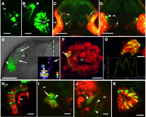
Expression of Rag1 in zebrafish olfactory system. (A) At 22 hours post-fertilization, a few olfactory sensory neurons express GFP under the Rag1 promoter. (B) At 3 dpf, fluorescing axons have reached the bulb. A single target (arrowhead) is innervated by brightly labelled axons. (C, D) Frontal view of an 8-day old larva, with DiI labeling of olfactory sensory neurons (red) and Bodipy labeling of inter-cellular spaces (dim green). (C) A shallow section of the labelled forebrain, with DiI-labelled olfactory axons visible (arrows). In a deeper section (D) the GFP-containing axons (arrowhead) can be seen, along with other DiI-labelled axons (arrow). (E) Dorsal view of an isolated forebrain from a 4 day-old fish, showing the left olfactory bulb. Anterior is to the top and the midline is indicated by the dotted line. Strong GFP fluorescence is seen in axon terminals in a single region of the lateral bulb (arrowhead), while axons with lower levels of GFP innervate other regions of the lateral bulb (arrows). The inset shows one optical section, colour-coded according to fluorescence intensity. Termini with high (arrowhead) and low (arrow) intensity are indicated. (F) Frontal view of glomerular structures in the olfactory bulb of a 4-day fish, labelled with an antibody to synaptic vesicles. Only one lateral structure is innervated by OSNs with strong GFP expression (arrow). Lateral is to the left, while dorsal is to the top. (G) A single optical section through the glomerular target containing GFP-expressing neurons. The marker for synaptic vesicles (red) and GFP appear to co-localize, as indicate by the linescan. (H) An olfactory pit labeled with DiI. The GFP-expressing cells (green) have not taken up DiI (red). (I, J) A Di8ANEPPQ-labeled olfactory system of a Rag1:GFP transgenic fish. In the olfactory bulb (I), some axons with strong GFP expression (green) are also labeled with Di8ANEPPQ (red; arrow), whereas others are not (arrowhead). (J) In this section through the olfactory epithelium, a GFP-expressing neuron was labeled with Di8ANEPPQ (arrow), whereas two others were not (arrowheads). (K) The olfactory pit of a fish transgenic for Rag1:GFP and omp:tauDsRed. GFP expressing cells (green) are distinct from those labeled with DsRed. Panels A, B, E, F and K are projections reconstructed from Z stacks. ob: olfactory bulb; op: olfactory pit. Bar = 20 µm (A, B, E, F, H, I, K); 50 µm (C, D,); 5 µm (G, J).
|
