
FIGURE
Fig. 4
- ID
- ZDB-FIG-050922-11
- Publication
- Barresi et al., 2005 - Hedgehog regulated Slit expression determines commissure and glial cell position in the zebrafish forebrain
- Other Figures
- All Figure Page
- Back to All Figure Page
Fig. 4
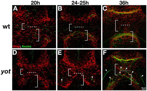
The post-optic glial bridge is disrupted in yot (gli2DR) mutants. Axons (anti-AT; green) and Gfap+ cells (red) at 20 hpf (A,D), 24-25 hpf (B,E), and 36 hpf (C,F) in wild type (WT; A-C) and yot (gli2DR) mutants (D-F). (D-F) POC and RGC axons do not cross the midline in yot (gli2DR) mutants (E,F, arrowheads) (Karlstrom et al., 1999). Gfap+ cells are reduced at the midline in the region where the POC would normally form (right brackets), and spread into the pre-optic area (left brackets). Frontal views, anterior up, dotted line marks the optic recess. |
Expression Data
| Gene: | |
|---|---|
| Fish: | |
| Anatomical Term: | |
| Stage Range: | 20-25 somites to Prim-25 |
Expression Detail
Antibody Labeling
Phenotype Data
Phenotype Detail
Acknowledgments
This image is the copyrighted work of the attributed author or publisher, and
ZFIN has permission only to display this image to its users.
Additional permissions should be obtained from the applicable author or publisher of the image.
Full text @ Development