
Fig. 3
- ID
- ZDB-FIG-050805-7
- Publication
- Gu et al., 2005 - Molecular cloning and expression of a novel CYP26 gene (cyp26d1) during zebrafish early development
- Other Figures
- All Figure Page
- Back to All Figure Page
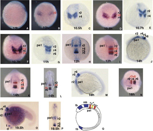
Expression pattern of cyp26d1 mRNA during segmentation period of zebrafish. (A, J, Q) Lateral view, dorsal right, anterior top; (B–I, K) dorsal view, dorsal forward, anterior top; (L, P) flatmount of anterior part of embryo; anterior top; (M, O) lateral view; anterior left, dorsal top; (N) dorsal view, dorsal forward, anterior left. cyp26d1—blue; krox20—red. (A) Expression of cyp26d1 gene is in presumptive hindbrain region at bud stage (10 hpf). (B) Double in situ hybridized embryos at bud stage with krox20 and cyp26d1 shows that the two separate dorsal regions of cyp26d1 expression are in presumptive r2–r4. (C) cyp26d1 mRNA is present in two stripes that are limited to lateral expression and do not meet at the midline of the anterior neuroectoderm at 2-somite stage (10.5 hpf). (D) Double in situ hybridization shows that zebrafish cyp26d1 is expressed in presumptive r2 and r4 and part of r3 region at 2-somite stage. (E–G) The lateral expression stripes at presumptive r2 merge at the midline of embryos at 2–3-somite stages (10.7–11 hpf) while the expression stripes at r4 do not. At 3-somite stage, cyp26d1 gene is also expressed in the stripes that are presumptive r6 and in pharyngeal arch (pa) one in addition to expression at r2 and r4 (G). (H, I) At 6-somite stage (12 hpf), the expression of cyp26d1 is in continuous bands at r2–r6 and also in pa1 with broad expression at r2 and high expression at r6. (J–P) The expression pattern is maintained from 10-somite stage (J–L), 18-somite stage (M, N) through 21-somite stage (19.5 hpf) (O, P) except that the expression level of the gene is greatly reduced at r2 and hardly detectable in r4 (J–P). At 21-somite stage, cyp26d1 is also found in a group of cells in telencephalon and diencephalons (O). (Q) A schematic diagram summarizes expression domains of the three cyp26 genes (cyp26a1, cyp26b1, and cyp26d1) in embryos at 3-somite stage. Blue areas show the expression regions of cyp26a1, red ones mark the expression regions of cyp26d1 and the yellow area is the region where cyp26b1 and cyp26d1 are co-expressed. d, Diencephalon; fb, forebrain; mb, midbrain; pa, pharyngeal arch; r, rhombomere; t, telencephalon; tb, tail bud. |
| Genes: | |
|---|---|
| Fish: | |
| Anatomical Terms: | |
| Stage Range: | Bud to 20-25 somites |
Reprinted from Gene expression patterns : GEP, 5(6), Gu, X., Xu, F., Wang, X., Gao, X., and Zhao, Q., Molecular cloning and expression of a novel CYP26 gene (cyp26d1) during zebrafish early development, 733-739, Copyright (2005) with permission from Elsevier. Full text @ Gene Expr. Patterns