
Fig. 1
- ID
- ZDB-FIG-050719-6
- Publication
- Zecchin et al., 2005 - Expression analysis of jagged genes in zebrafish embryos
- Other Figures
- All Figure Page
- Back to All Figure Page
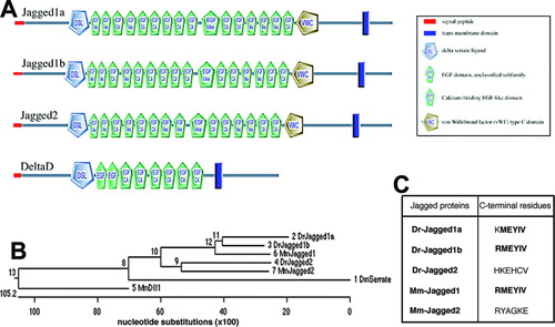
Domain structure and phylogenetic relationships of zebrafish Jagged proteins. A: Schematic diagram of the motif/domain structure of zebrafish Jagged1a, Jagged1b, Jagged2, and DeltaD, obtained with the SMART program (http://smart.embl-heidelberg.de). The cysteine-rich domain encompasses the von Willebrand factor type C-like domain. B: Dendrogram of zebrafish (Danio rerio, Dr) Jagged1a, Jagged1b, and Jagged2; mouse (Mus musculus, Mm) Jagged1, Jagged2, and Dll1 (Delta-like1); and Drosophila (Drosophila melanogaster, Dm) Serrate, obtained with the MegAlign program that uses the Clustal W algorithm (Thompson et al., [1994]). C: C-terminal hexapeptides of zebrafish (Dr) and mouse (Mm) Jagged proteins. The hexapeptide of Jagged1b is identical, whereas that of Jagged1a differs by one amino acid, from the sequence of mouse Jagged1, shown to represent a functional PDZ-ligand (Hock et al., [1998]; Ascano et al., [2003]). The conserved amino acids in the three hexapeptides are in bold characters. The hexapeptides of zebrafish and mouse Jagged2 are not conserved and neither resemble a PDZ-ligand (Hock et al., [1998]; Ascano et al., [2003]). |