|
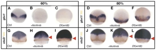
gbx1 and otx2 expression in Wnt8 loss-of-function experiments. (A-C) Embryos stained for gbx1 and (D-F) embryos stained for gbx1 and Ntl protein (brown). (A-C) Control embryo (A), embryo injected with MoWnt8 (B) and Df(Wnt8) mutant embryos (C) at 60%. gbx1 is not expressed at 60% in absence of Wnt8. (D-F) Control embryo (D), embryo injected with MoWnt8 (E) and Df(Wnt8) mutant embryo (F) at 80%. gbx1 expression is observed at 80% onwards and overlaps partially with the Ntl domain. (G-L) Embryos stained for otx2 and Ntl protein (brown). (G-I) Control embryos (G), embryo injected with MoWnt8 (H) and Df(Wnt8) mutant embryo (I) at 60%. (J-L) Control embryos (J), embryos injected with MoWnt8 (K) and Df(Wnt8) mutant embryo (L) at 80%. Laterally a posterior shift of the otx2 expression domain is visible (red arrowheads) in the morphants and in the Df(Wnt8) mutant embryos. (A-L) Dorsal views, anterior upwards.
|