
Fig. 4
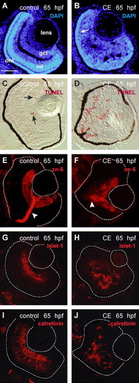
Differentiation and laminar organization of the retina in control and γ-secretase-inhibited (CE) embryos at 65 hpf. (A, B) Retinal cell nuclei labeled by DAPI (blue). The retinal cell layers are distinguishable in the control retina (A) but in the CE-treated retina only the onl (arrow) can be identified (B). TUNEL-labeled control (C) and CE-treated (D) retinal cryosections (Fast Red—red). The arrows in (C) point to apoptotic cells in the wild-type retina. Ganglion cells and their axons in the optic nerve (arrowhead) were labeled by zn-5/neurolin in control (E) and CE-treated (F) retinas (N = 12 embryos). Ganglion cell nuclei were labeled by islet-1 antibody in control (G) and CE-treated (H) retinas (N = 10 embryos). The retinal boundary is outlined. The calretinin antibody labeled ganglion, amacrine, and bipolar cells (based on location and morphology) in the retinas of control embryos (I) and neurons in the CE-treated embryos (J) (N = 8 embryos). Scale bar = 50 μm (A–J). Abbreviations as in Fig. 1. |
| Genes: | |
|---|---|
| Fish: | |
| Condition: | |
| Anatomical Terms: | |
| Stage: | Pec-fin |
Reprinted from Developmental Biology, 278(2), Bernardos, R.L., Lentz, S.I., Wolfe, M.S., and Raymond, P.A., Notch-Delta signaling is required for spatial patterning and Muller glia differentiation in the zebrafish retina, 381-395, Copyright (2005) with permission from Elsevier. Full text @ Dev. Biol.