
Fig. 6
- ID
- ZDB-FIG-050304-4
- Publication
- Jackman et al., 2004 - Fgf signaling is required for zebrafish tooth development
- Other Figures
- All Figure Page
- Back to All Figure Page
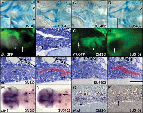
SU5402 inhibits zebrafish pharyngeal tooth morphogenesis. (A–D) Ventral views of 82-h cartilage-stained specimens centered on the posterior pharynx and focused at the level of the teeth and ceratobranchial cartilages, anterior to the left. (A) Control larvae treated from 32 to 82 h with 0.5% DMSO develop normal pharyngeal cartilages and teeth (arrowheads and inset). (B) Larvae treated from 32 to 82 h with 25 μM SU5402 exhibit pharyngeal cartilage reduction, and teeth are absent. However, occasionally, specimens are seen in these treatments with more severe cartilage reductions (C), or with more normal cartilages and teeth present (arrowhead and inset, D). (E) A fluorescent/bright-field double image of a 78-h tooth germ in the fli1:GFP transgenic line. Non-GFP-expressing dental epithelium (mineralized portion of tooth indicated with arrow) surrounds GFP-expressing dental mesenchyme (arrowhead). (F) A toluidine blue-stained sagittal section at 72 h showing the dental mesenchyme (arrowhead), mineralized tooth tip (arrow), and the location of the 6th pharyngeal pouch (double-arrow). (G) In a control larva treated with 0.5% DMSO from 32 to 78 h, the GFP-expressing dental mesenchyme (arrowhead) surrounded by non-expressing epithelium is located caudad to the 6th pouch (double-arrow). (H) In 32–78 h SU5402-treated individuals, the 6th pharyngeal pouch has adopted a rounded morphology (double-arrow), and no gap in GFP expression is detectable in the region where the tooth would normally form (arrowhead). (I) Toluidine blue stained transverse section of the tooth germ after DMSO exposure from 32 to 56 h. The pharyngeal epithelium is colored in red in (J), with the curved dental epithelium visible (arrow). (K and L) After 32–56 h SU5402 treatment, morphogenesis of the dental epithelium is no longer apparent, although this epithelium may be slightly thickened (arrow). (M and N) Expression of pitx2 in the tooth germs is identical between 32–56 h DMSO-treated controls (arrow, M) and 32–56 h SU5402-treated individuals (arrow, N). Dorsal views, anterior to the left. (O) Likewise, transverse sections reveal normal pitx2 expression and dental epithelial morphogenesis at 56 h in DMSO control embryos (arrow), while in 32-56 h SU5402-treated specimens (P), pitx2 expression remains in the pharyngeal epithelium but no epithelial morphogenesis is visible (arrow). Scale bars = 100 μM. |
| Gene: | |
|---|---|
| Fish: | |
| Condition: | |
| Anatomical Terms: | |
| Stage: | Long-pec |
Reprinted from Developmental Biology, 274(1), Jackman, W.R., Draper, B.W., and Stock, D.W., Fgf signaling is required for zebrafish tooth development, 139-157, Copyright (2004) with permission from Elsevier. Full text @ Dev. Biol.