
Fig. 2
- ID
- ZDB-FIG-050214-2
- Publication
- Jackman et al., 2004 - Fgf signaling is required for zebrafish tooth development
- Other Figures
- All Figure Page
- Back to All Figure Page
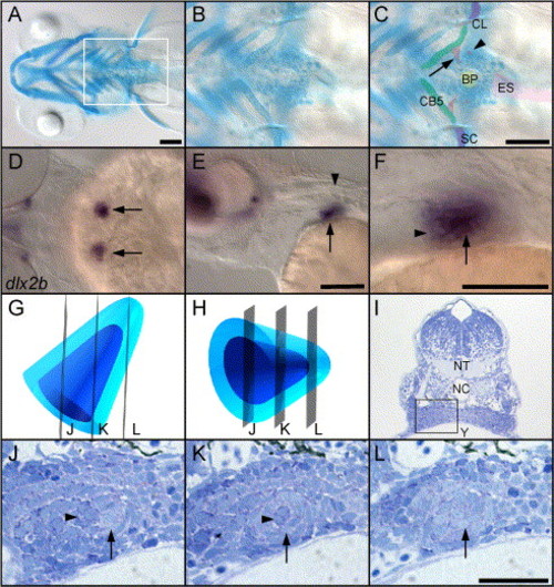
Zebrafish teeth are located deep in the posterior, ventral portion of the pharynx, but can be visualized by several methods. (A–C) Ventral views of 5 day larvae stained with alcian green to label cartilages and highlight mineralized teeth, anterior to the left. (A) A broad, ventral view of the head shows the area where teeth develop (box). (B and C) Unlabeled and labeled views of the posterior pharynx. The earliest-formed teeth (arrow) are by this time attached to the 5th ceratobranchial cartilage (CB5), while the third-formed teeth (arrowhead) have just begun to mineralize and are not yet attached. The second tooth pair is out of the plane of focus. Elements of the pectoral girdle flank the tooth-forming region (scapulocoracoid, SC; cleithrum, CL), the esophagus lies caudally (ES), and the keratinized bite pad (carpstone), against which the teeth ultimately bite, is present in the midline on the dorsal surface of the pharynx (BP). (D–F) Expression of dlx2b at 56 h reveals the location and orientation of the first pair of tooth germs, before they have begun to mineralize. Anterior to the left. (D) Dorsal view focused through the hindbrain, arrows indicate the tooth germs. (E) Lateral view shows a tooth germ (arrow) just ventral and rostral to the first myotome (arrowhead). (F) Close-up of (E) reveals that the dental epithelium (arrow) surrounds the dental mesenchyme except rostrally (arrowhead). (G and H) Diagrams of 56 h tooth germs with the dental mesenchyme colored dark blue, and the dental mesenchyme a lighter shade. (G) Tooth germ viewed from dorsal, mimicking the orientation of the left-side tooth germ in (D). Transverse planes of section are indicated (J–L). (H) Tooth germ viewed laterally as in (E) and (F). (I) Transverse section of a 56-h larvae at the level of the developing teeth to indicate the location of the left-side tooth germ in this plane of section (box; NT, neural tube; NC, notochord; Y, yolk; up is dorsal). (J–L) Three typical shapes of a 56-h tooth germ in transverse section. (J) At the rostral end, a group of dental mesenchyme cells (dark blue, arrowhead) is flanked medially by a crescent of dental epithelium (light blue, arrow). (K) In the center of the tooth germ, the epithelium (arrow) completely surrounds a core of mesenchyme (arrowhead). (L) Caudally, the section does not pass through the mesenchyme at all, and only dental epithelial cells are seen (arrow). Scale bars = 100 μm. |
| Gene: | |
|---|---|
| Fish: | |
| Anatomical Term: | |
| Stage: | Long-pec |
Reprinted from Developmental Biology, 274(1), Jackman, W.R., Draper, B.W., and Stock, D.W., Fgf signaling is required for zebrafish tooth development, 139-157, Copyright (2004) with permission from Elsevier. Full text @ Dev. Biol.