|
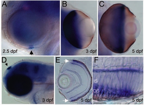
Temporal expression pattern of pcdh15b in differentiating photoreceptor cells. (A) At 2.5 dpf, pcdh15b is expressed in the first differentiating photoreceptors at the ventral margin of the eye (arrowhead). (B) At 3 dpf, the whole proximal optic cup is stained. (C) At 5 dpf, expression is only present at the margin of the optic cup, no expression is visible in the proximal eye. (D) Expression in the epiphysis (arrow) and brain at 3 dpf. pcdh15b expression in the eye is specific to photoreceptor cells (E,F; cryosection of a larva stained as in C) Arrows in (E) indicate photoreceptor proliferation zones shown at higher magnification in (F). Scale bar in F, 50 µm for A; 40 µm for B,C,E; 150 µm for D; and 5 µm for F. IS, inner segment; OS, outer segment.
|