
Fig. 6
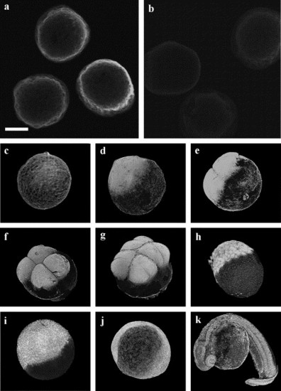
Immunofluorescence localization of Yes kinase in the zebrafish embryo. Chorion-free zygotes were fixed at various times after fertilization, then stained with the anti-yes antibody, followed by FITC-conjugated goat anti-rabbit IgG. Examination by confocal fluorescence microscopy utilizing a 24-μm optical section demonstrates localization of the anti-Yes antibody to the cortex of the 2.5-min zygote (a), while staining by the preimmune serum was negative (b). Images presented in panels c–k are three-dimensional reconstructions made from optical sections of embryos fixed at 2.5 min (c) and 30 min postinsemination (d), two cell stage (e), four cell stage (f), eight cell stage (g), blastula (h), gastrula (i), early segmentation stage (j), and pharyngula (k). Scale bar = 300 μm. |
| Gene: | |
|---|---|
| Fish: | |
| Anatomical Term: | |
| Stage Range: | 1-cell to Prim-5 |
Reprinted from Developmental Biology, 277(1), Tsai, W.B., Zhang, X., Sharma, D., Wu, W., and Kinsey, W.H., Role of Yes kinase during early zebrafish development, 129-141, Copyright (2005) with permission from Elsevier. Full text @ Dev. Biol.