
- Title
-
Role of Yes kinase during early zebrafish development
- Authors
- Tsai, W.B., Zhang, X., Sharma, D., Wu, W., and Kinsey, W.H.
- Source
- Full text @ Dev. Biol.
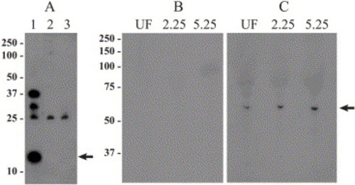
Detection of the Yes kinase in zebrafish embryos. The specificity of the anti-Yes antibody was tested by Western blot (panel A) of GST fusion proteins encoding the U + SH3 domain of zebrafish Yes (lane 1), the U + SH3 domain of Xenopus Src (lane 2), or the U + SH3 domain of zebrafish Fyn (lane 3). Fusion proteins were digested with thrombin to release the GST component, then resolved by SDS-PAGE, and blots were probed with the anti-Yes antibody. As seen in panel A, the antibody recognized the 13-kDa U + SH3 insert only in the sample from zebrafish Yes (lane 1, arrow). Higher MW bands in lane A represent partial digestion products containing Yes sequence. Some staining of the 25-kDa GST band was present in all samples, indicating that some anti-GST antibodies still remain in the partially purified antibody. The expression of Yes in eggs and early embryos was tested by Western blot of unfertilized eggs (UF), blastula obtained at 2.25 hpf, and early gastrula (50% epiboly) obtained at 5.25 hpf. Each lane was loaded with the equivalent of two embryos represented approximate 130 μg protein. The blots were probed with preimmune serum (panel B) or the partially purified antibody (panel C). The position of the 62-kDa Yes protein is indicated by an arrow at right. |
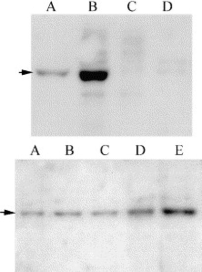
Detection of Yes kinase activity in zebrafish embryos. Detergent extracts prepared from 100 embryos collected at 5.25 h postinsemination were incubated with control serum (A) or the partially purified anti-Yes antibody (B) at 1:250 dilution. Immune complexes were collected with protein A-Sepharose, washed, and incubated in a kinase reaction as described in Materials and methods. The reaction products were resolved by SDS-PAGE, treated with 1N KOH to reduce contamination by 32P-Ser and 32P-Thr, then the phosphoproteins were detected by autoradiography. The anti-Yes antibody immunoprecipitated kinase activity evident as a 62-kDa band (arrow). An additional protein of 110–120 kDa was also phosphorylated in these reactions. Autophosphorylation assays of immunoprecipitates prepared at different times during development demonstrated that Yes kinase activity was detected in the unfertilized egg (C), at 3.5 h (D), and 5.25 h post fertilization (E). |
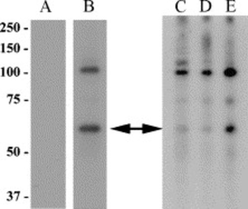
Detection and phosphorylation of phosphorylated FAK in zebrafish embryos. Co-immunoprecipitation and phosphorylation of FAK in Yes immunoprecipitates (upper panel) was detected by preparing immunoprecipitates from pooled membrane fractions (1000 embryos) collected 6 h postfertilization with either anti-Yes antibody at 5 μg/ml (A and B) or anti-Fyn3 antibody at 1 μg/ml (C and D). The immune complexes were washed and incubated in kinase buffer containing 50 μM Na3VO4 to suppress phosphatase activity. ATP was added to some samples at a final concentration of 100 μM (B and D) to initiate phosphorylation of FAK, and all samples were incubated at 25°C for 5 min, then heated in SDS-PAGE sample buffer to stop the reaction. Samples were resolved by SDS-PAGE and blotted to immobilon. Blocking solution for this experiment consisted of 5% BSA in TTBS containing 50 μM Na3VO4,, and the presence of FAK phosphorylated on Tyr 861 was detected using the anti-FAK[pY861] antibody at 0.5 μg/ml, followed by anti-rabbit-HRP, and chemiluminescence detection. The phosphorylated FAK is apparent as a single band indicated by the arrow. The phosphorylation of FAK at Tyr 861 in vivo (lower panel) was detected by Western blot analysis of pooled membrane fractions (50 μg protein) prepared at 2.5 hpf (A), 3.5 hpf (B), 4 hpf (C), 5.25 hpf (D), and 6.5 hpf (E). |
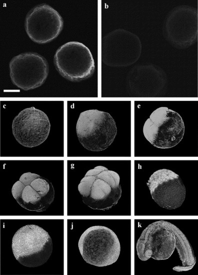
Immunofluorescence localization of Yes kinase in the zebrafish embryo. Chorion-free zygotes were fixed at various times after fertilization, then stained with the anti-yes antibody, followed by FITC-conjugated goat anti-rabbit IgG. Examination by confocal fluorescence microscopy utilizing a 24-μm optical section demonstrates localization of the anti-Yes antibody to the cortex of the 2.5-min zygote (a), while staining by the preimmune serum was negative (b). Images presented in panels c–k are three-dimensional reconstructions made from optical sections of embryos fixed at 2.5 min (c) and 30 min postinsemination (d), two cell stage (e), four cell stage (f), eight cell stage (g), blastula (h), gastrula (i), early segmentation stage (j), and pharyngula (k). Scale bar = 300 μm. |
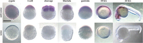
Localization of c-Yes transcripts in the zebrafish embryo. c-Yes mRNA was localized by hybridization with a digoxigenin-labeled antisense riboprobe, or with the complimentary sense probe as a control. Bound probe was localized with anti-digoxigenin-phosphatase. Scale bar = 300 μm. |
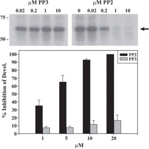
PP2 inhibition of Yes kinase activity, effect on development. The effectiveness of PP2 on Yes kinase was tested by immune complex assay (upper panel). Yes immunoprecipitates from 5-h zebrafish embryos were incubated for 5 min with different concentrations of PP2 or PP3 (control). Protein tyrosine kinase activity in the immunoprecipitates was then measured by an autophosphorylation reaction in the presence of [γ-32P]ATP, and the phosphorylated proteins were resolved by SDS-PAGE and detected by autoradiography. The position of the 62-kDa band representing Yes kinase is indicated by the arrow. The effect of PP2 on development (lower panel) was tested by culturing zebrafish zygotes in different concentrations of PP2 and PP3. After 5 h incubation, the embryos were washed free of the inhibitors. At 5.5 h of development, groups of 25–30 embryos were examined and those scored as having successfully undergone 50% epiboly were classified as having developed normally. Values represent the average of three experiments ± S.E.M. |
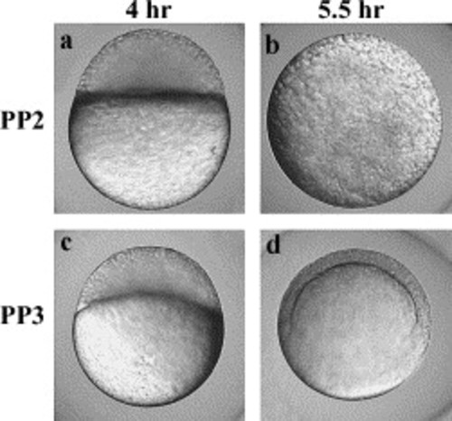
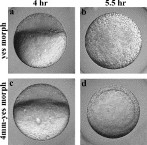
Morphology of the PP2-treated embryos. Fertilized eggs (3 min postinsemination) were treated with 4 μM PP2, or PP3 as a control for 5 h. Embryos resulting from eggs treated with PP2 exhibited normal morphology at the high/sphere stage (a) but they exhibited a range of defects involving failure of epiboly (b). Embryos treated with PP3 exhibited normal development (c–d). |
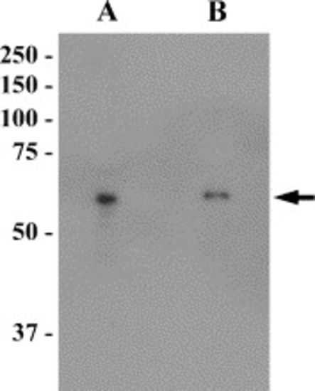
Effect of control and Yes antisense morpholino on the expression of Yes kinase. Embryos resulting from zygotes injected with 12.5 ng of fluorescein tagged control or Yes antisense morpholino 5.5 h were homogenized and analyzed by Western blot. The Yes protein was evident as a band at 62 kDa (arrow). Densitometric scanning of the band in this experiment revealed that the injection of Yes antisense morpholino reduced Yes protein to a level of 62% of that in the control. EXPRESSION / LABELING:
|
Effect of yes antisense morpholino oligonucleotide injection on development. Embryos resulting from zygotes injected with 12.5 ng of the Yes antisense oligonucleotide exhibit normal development up to the sphere stage (a), but epiboly was defective and the normal embryonic germ layers were not formed (b). Embryos resulting from zygotes injected with 12.5 ng of the mutated control Yes antisense oligonucleotide (c–d) gastrulated normally. Scale bar = 300 μm. |
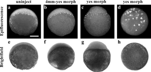
Effect of yes antisense morpholino oligonucleotide injection on distribution of embryonic cells. Embryos resulting from control, uninjected eggs (a and e), or from eggs injected with 12.5 ng of the control (4-mm yes morph; b and f) and Yes antisense oligonucleotide (c, d, g, and h) were fixed at 5.5 hpf (50% epiboly) and stained with DAPI to visualize cell nuclei. Bright-field images (panels e–h) show the overall morphology of the embryo, while fluorescence micrographs (panels a–d) show the distribution of cell nuclei over the embryo. Scale bar = 200 μm. |
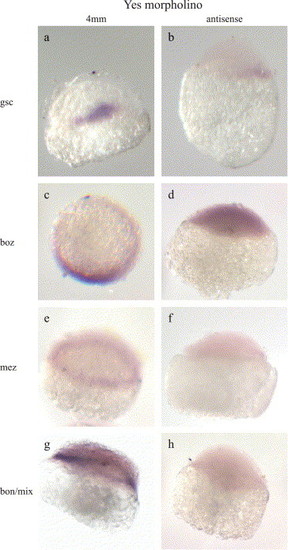
Effect of antisense morpholino on the spatial localization of goosecoid, boz, mezzo, and bon/mixer transcripts. Embryos resulting from zygotes injected with 12.5 ng of the control 4-mm yes morpholino control (panels a, c, e, and g) or with yes antisense morpholino (b, d, f, and h) were fixed after 5.5 h of development (50% epiboly). Transcripts were localized by in situ hybridization as described in Materials and methods. Scale bar = 300 μm. EXPRESSION / LABELING:
|
Reprinted from Developmental Biology, 277(1), Tsai, W.B., Zhang, X., Sharma, D., Wu, W., and Kinsey, W.H., Role of Yes kinase during early zebrafish development, 129-141, Copyright (2005) with permission from Elsevier. Full text @ Dev. Biol.