
- Title
-
her4, a zebrafish homologue of the Drosophila neurogenic gene E(spl), is a target of notch signalling
- Authors
- Takke, C., Dornseifer, P., von Weizsacker, E., and Campos-Ortega, J.A.
- Source
- Full text @ Development
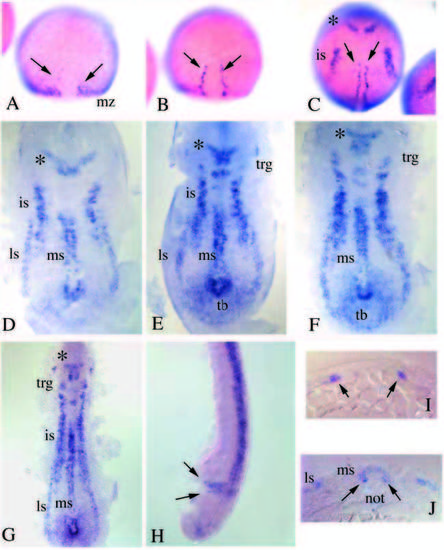
Distribution of her4 transcripts revealed by whole-mount in situ hybridization. (A) 70% epiboly. her4 expression appears in the epibolic margin, the midline is devoid of transcript. (B) 90% epiboly. A row of hypoblastic her4-expressing cells is visible on either side of the midline (arrows). (C) 100% epiboly. her4 expression in two lateral stripes in the neural plate (is) and in a V-shaped expression domain (asterisk) in the prospective brain region; the hypoblastic expression domains elongate anteriorly. (D) Flat preparation of a tailbud-stage embryo. Expression domains in the neural plate comprise two lateral stripes, one in an intermediate position (is) and one further lateral and caudal (ls), which will fuse during later stages of development; in addition there is a medial stripe in the neural plate which overlies the hypoblastic expression domain. (E) 1- somite stage. The intermediate and lateral expression domains are still distinct. (F) 2- somite stage. Intermediate and lateral expression domains have fused and the medial stripe has elongated. her4 transcription in the primordium of the trigeminal ganglion (trg) becomes visible. (G) 5-somite stage. (H) 24 h embryo. her4 transcription is detectable in two bands in the presomitic mesoderm. (I) Transverse section through the 90% epiboly embryo shown in B, which reveals the hypoblastic expression (arrows). (J) Transverse section through a 6-somite embryo, showing the lateral and medial expression domains in the neural plate, and the hypoblastic expression in the adaxial mesoderm. |
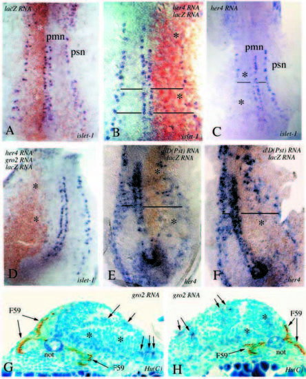
(A-F) Flat preparations of embryos labelled by in situ hybridization with an islet-1 probe (blue) and stained with an antibody against β-galactosidase (brown). pmn primary motoneurons, psn primary sensory neurons. Asterisks in all panels indicate the affected side. (A) 4-somite stage control embryo injected with lacZ RNA alone. (B-C) 4-somite stage embryos injected with full-length her4 and lacZ RNA. Both embryos have been stained for islet-1 expression, the one in B, in addition, for β-galactosidase expression. A reduction in the number of of islet-1-expressing cells can be observed on one side. The horizontal lines in these and the other panels indicate the extent of the enlargement of the neural plate on the affected side. (D) A 4-somite stage embryo injected with full-length her4, groucho2 and lacZ RNA, stained for islet-1 and β- galactosidase expression. Note the enlargement of the neural plate and the complete lack of islet-1-positive cells on the injected side. (E,F) Two 4-somite stage embryos which have been injected with deltaD(Pst) and lacZ RNA, and stained for her-4 and β-galactosidase expression; the anti-β-galactosidase staining was kept to a minimum in this case, to avoid brown overstaining. Notice that her-4 expression is reduced on the brown side. (G,H) Cross sections of 24 h embryos injected with groucho2 RNA and stained with two antibodies: F59, which recognises the myosin heavy chain (Miller et al., 1989) and labels the adaxial mesodermal cells and their derivatives (brown product; see Devoto et al., 1996), and Hu(C), a neuronal marker (Kim et al., 1996). Notice the asymmetry of the neural tube (not: notochord), one side of which is much larger. Hu(C) cells (blue product) are indicated with arrows on the affected side. Notice that mesodermal development, as judged by the F59 staining, is also affected on the same side. |
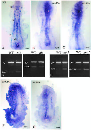
Distribution of her4 transcripts in the wild-type (A) and following injection of nic RNA. her4 transcription is activated in the neural plate (asterisks, B,C). RT-PCR analysis of target gene expression following misexpression of nic (D) and ngn1 (E). As an internal standard a fragment of the elongation factor 4a (EF) was amplified. Activation of the NOTCH pathway by misexpression of nic leads to reduction of deltaD transcription and activation of her4 transcription, whereas misexpression of ngn1 activates transcription of both deltaD and her4. (F,G) her4 in situ hybridizations following her4 and nic RNA injections. Both were processed simultaneously, under identical conditions. The density of her4 transcripts in F is much higher than in G. |
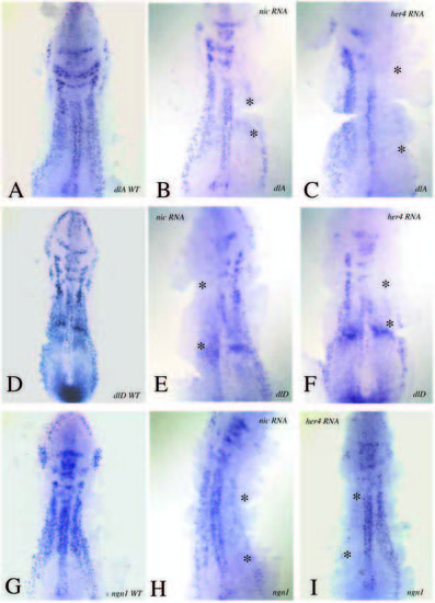
Wild-type expression patterns of deltaA (A), deltaD (D) and ngn1 (G) in 5-somite stage embryos as revealed by in situ hybridization. (B,E,H) Embryos of the same age injected with nic RNA and scored for expression of deltaA (B), deltaD (E) and ngn1 (H). Transcription of these genes is reduced on the injected side of the embryo (asterisks). (C,F,I) Embryos injected with full-length her4deltaA (C), deltaD (F) and ngn1 (I). Transcription of these genes is reduced (asterisks). |

Flat preparations of 6-somite stage embryos injected with deltaD(Pst) RNA and stained for islet-1 expression by in situ hybridization (A-C). Arrows and asterisks label the affected side. (D,E) 1-somite stage embryos injected with deltaD(Pst) RNA and scored for ngn1 transcription, detected by in situ hybridization with a ngn1 probe (D) and by double staining for β-galactosidase (E). (F,G) 6- and 8- somite stage embryos, treated as in D and E. Inhibition of DELTA signalling leads to an activation of ngn1 transcription within the limits of its normal expression stripes. |
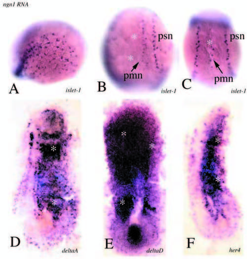
(A-C) 4-somite stage embryos injected with ngn1 RNA and stained for islet-1 expression by in situ hybridization. (A) Lateral view of an injected embryo showing ectopic islet-1-positive cells in the ectoderm adjacent to the neural plate. (B,C) Dorsal views reveal a reduction in the number of islet-1-positive cells within the neural plate following misexpression of ngn1 (asterisks). (D,E,F) 3-somite stage embryos injected with neurogenin1 RNA and stained for the expression of deltaA (D), deltaD (E) and her4 (F). Transcription is activated within the neural plate (asterisks). |