
- Title
-
Zebrafish ultraviolet visual pigment: absorption spectrum, sequence, and localization
- Authors
- Robinson, J., Schmitt, E.A., Hárosi, F.I., Reece, R.J., and Dowling, J.E.
- Source
- Full text @ Proc. Natl. Acad. Sci. USA
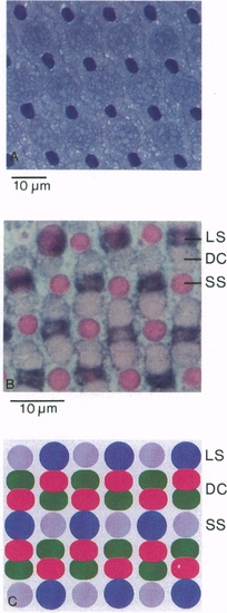
(Top) Light micrograph of zebrafish retinal mosaic. This section is cut through the densely stained outer segments of the short single photoreceptors and shows their arrangement. (1% methylene blue/1% azure blue). (Middle) Zebrafish cone mosaic: A 5-μm section stained with eosin and an antisense riboprobe to blue opsin. LS, long single cone; DC, double cone; SS, short single cone. The section is cut through the inner segments of the long single and double cones and through the outer segments of the short single cones. (Bottom) Representation of the retinal mosaic in zebrafish. The cones are arranged in rows, resulting in a regular mosaic arrangement. Long single (blue-sensitive) cones are represented by the blue circles; double cones, by the red and green ovals; and the short single cones, by the violet circles. |
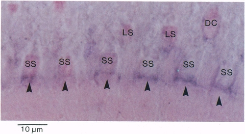
Longitudinal section (5 μm) of a dark-adapted zebrafish photoreceptor layer, hybridized to a DIG-labeled riboprobe generated antisense to ZFO2. mRNA present in the myoids of all short single photoreceptors (SSC) hybridized to this probe. No signal was detected in either member of the double cones (DC) or in the long single cells (LS). Furthermore, no hybridization to rods was detected. (Bar = 10 μm.) |