
Image
|
Figure Caption
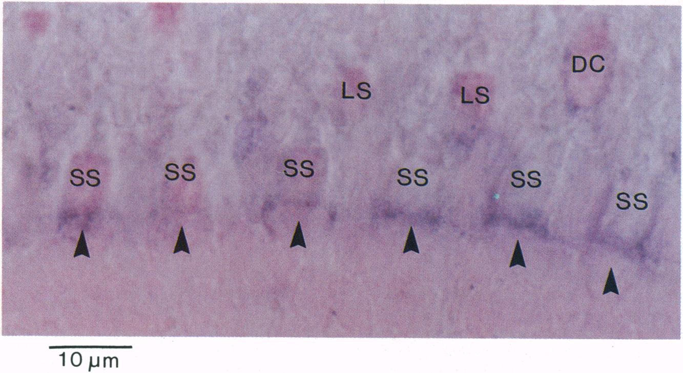
Fig. 4 Longitudinal section (5 μm) of a dark-adapted zebrafish photoreceptor layer, hybridized to a DIG-labeled riboprobe generated antisense to ZFO2. mRNA present in the myoids of all short single photoreceptors (SSC) hybridized to this probe. No signal was detected in either member of the double cones (DC) or in the long single cells (LS). Furthermore, no hybridization to rods was detected. (Bar = 10 μm.)
Acknowledgments
This image is the copyrighted work of the attributed author or publisher, and
ZFIN has permission only to display this image to its users.
Additional permissions should be obtained from the applicable author or publisher of the image.
Full text @ Proc. Natl. Acad. Sci. USA