
- Title
-
Retinoic acid establishes ventral retinal characteristics
- Authors
- Hyatt, G.A., Schmitt, E.A., Marsh-Armstrong, N., McCaffery, P., Dräger, U.C., and Dowling, J.E.
- Source
- Full text @ Development

The level and orientation of transverse sections for control and RA treated embryos are indicated by lines A, B and C, respectively in D. Note the optic stalks (OS) which extend outward from the forebrain are located at the anterior of the eye cups at 24 hpf in both control and RAtreated embryos. A boundary (b), an extension of the optic lumen, divides the dorsal and ventral retinas of RA-treated embryos. (A) The eyecup of a control embryo at 30 hours postfertilization (hpf) with a single pseudostratified retina (R) and lens (L). (B) At 24 hpf, RA-treatment induced an apparent duplicated retina with a dorsal (D) and ventral (V) retina of slightly reduced size. (C) A more anterior section through the duplicated retina along the length of the optic stalk (OS) which extends laterally from the forebrain at 24 hpf (line C in D). The dorsal half of the stalk (Dos) is associated with the dorsal retina (D) whereas the ventral half of the optic stalk (Vos) is continuous with a layer of pigment epithelium (small arrows) which extends along the ventral retina (V). Note that the optic lumen extends between the dorsal and ventral retinal epithelia which creates a boundary (b) between them at the midline of the eyecup. D-V, dorsal-ventral axis; FB, forebrain; y, yolk; bar, 40 µm. PHENOTYPE:
|

(A) A control embryo (top) and embryo treated with RA after the critical period (bottom) is shown schematically at 36 hpf. In controls the choroid fissure (cf) is located at the ventral pole of the eyecup. In RA-treated embryos, the optic stalk (OS) and choroid fissure (cf) remains positioned at the anterior of the eyecup due to lack of eye rotation. Lines B, C and D indicate the orientation and level of transverse and horizontal sections shown in B, C and D, respectively. (B) A transverse section along the dorsal-ventral axis (D-V) at 36 hpf after RA treatment between the 7- and 9-somite stages (i.e. shortly after the critical period). A small retinal epithelium (asterisk) lies ventral to the original retina (R) and lens (L). As found in embryos treated during the critical period, an extension of the optic lumen (white arrows) divides the two retinal epithelial layers and the pigment epithelium (pe) extends across the boundary. The lens (L) remains associated with the ectoderm which is unusually thick (arrows). Bar, 40 mm. (C,D) Sections through the midline of the eyecups at 36 hpf in a control embryo (C) and an embryo treated with RA after the critical period (D). (C) In horizontal sections of controls, a single retina (R) and lens (L) are observed and ventricles (V) are well formed within regions of the forebrain (Fb). Mitotic figures (white arrows) occur along the optic lumen adjacent to the pigment epithelium. The optic stalks which have atrophied are not evident as they lie ventral to the eycup midline at 36 hpf. (D) An embryo at 36 hpf after a 2 hour RA treatment between the 7- and 9-somite stages. Sections through the midline of the eyecup show the optic stalks (OS) are more than 30 mm wide and extend from the anterior region of the eyecups across the anterior surface of the forebrain. Distortions in the shape of the eyecups and lenses (black arrows) are observed. Numerous mitotic profiles (white arrows), associated with the partial retinal epithelial layer (see Fig. 2B), are located throughout the deeper regions of the retina (R) in addition to those along the optic lumen (arrowheads). The ventricles within the forebrain are not observed. A-P, anteriorposterior axis; y, yolk; bar, 60 µm. PHENOTYPE:
|

(A-D) A lateral view of control embryos (A,B) and RAtreated embryos (C,D) showing localization of pax[b] mRNA expression by whole-mount in situ hybridization. (A) An 18 hour control embryo with localization of pax[b] transcripts within the anterior/ventral region the eye adjacent to the optic stalk (os). (B) At 24 hpf, pax[b] transcripts in control embryos have been restricted to the optic stalk and within the choroid fissure (cf). (C) At 18 hpf, a ventral-to-dorsal spread of pax[b] transcripts is observed in RAtreated embryos. (D) By 24 hpf, pax[b] is expressed throughout the dorsal and ventral regions of the eyecups (double arrow) in treated embryos (arrow). (E) Diagrams of embryos shown above illustrates changes in pax[b] expression in control and RA-treated embryos. D-V, dorsal-ventral axis; y, yolk; bar, 210 µm. EXPRESSION / LABELING:
|
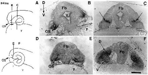
The localization of pax[b] transcripts at 24 hpf in transverse sections (5 µm) of eyes from control embryos (B,C) and embryos treated with all-trans RA (E,F). (A) Schematic of a control embryo at 24 hpf indicating the level of transverse sections (lines B and C) shown in B and C, respectively. (B,C) In control embryos, transcripts are localized to the optic stalks (os in B) and to a sharp band extending across the retina (arrows in C) in the anterior region of the eyecup. (D) Schematic of an RA-treated embryo at 24 hpf illustrating the level of transverse sections (lines E and F) shown in E and F. (E,F) In treated embryos, pax[b] expression is present within the optic stalks (OS in E) and throughout the neuroepithelium of both the dorsal (D) and ventral (V) retina of the duplication. D-V, dorsal-ventral axis; y, yolk; bar, 65 µm in B, bar, 80 µm in C, E and F. EXPRESSION / LABELING:
|

In situ hybridization of msh[c] transcripts at 20 hpf in control embryos (A,D) and embryos treated with RA during the critical period (B). (A) In control embryos, msh[c] expression is localized to a small patch in the dorsal/posterior region of the eyecup (arrow) at 20 hpf. (B) No msh[c] expression is observed in the eyes (e) of treated embryos. (C) Illustration of a 20 hpf control embryo indicating the level of the transverse section (line D) shown in D. The arrow indicates the patch of msh[c] expression shown in A. (D) In transverse section, msh[c] expression (arrow) is found in the dorsal region of the neuroepithelium of the retina (R) in sections through the posterior region in the eyes of control embryos. D-V, dorsal-ventral axis; A-P, anterior-posterior axis; bar in A and B, 180 µm; bar in D, 50 µm. EXPRESSION / LABELING:
|