
|
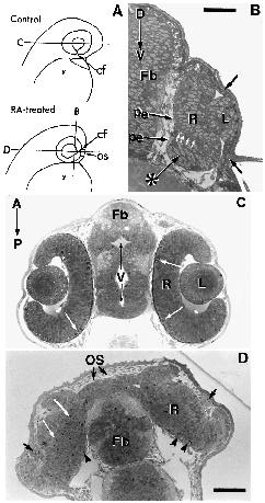
Fig. 2
(A) A control embryo (top) and embryo treated with RA after the critical period (bottom) is shown schematically at 36 hpf. In controls the choroid fissure (cf) is located at the ventral pole of the eyecup. In RA-treated embryos, the optic stalk (OS) and choroid fissure (cf) remains positioned at the anterior of the eyecup due to lack of eye rotation. Lines B, C and D indicate the orientation and level of transverse and horizontal sections shown in B, C and D, respectively. (B) A transverse section along the dorsal-ventral axis (D-V) at 36 hpf after RA treatment between the 7- and 9-somite stages (i.e. shortly after the critical period). A small retinal epithelium (asterisk) lies ventral to the original retina (R) and lens (L). As found in embryos treated during the critical period, an extension of the optic lumen (white arrows) divides the two retinal epithelial layers and the pigment epithelium (pe) extends across the boundary. The lens (L) remains associated with the ectoderm which is unusually thick (arrows). Bar, 40 mm. (C,D) Sections through the midline of the eyecups at 36 hpf in a control embryo (C) and an embryo treated with RA after the critical period (D). (C) In horizontal sections of controls, a single retina (R) and lens (L) are observed and ventricles (V) are well formed within regions of the forebrain (Fb). Mitotic figures (white arrows) occur along the optic lumen adjacent to the pigment epithelium. The optic stalks which have atrophied are not evident as they lie ventral to the eycup midline at 36 hpf. (D) An embryo at 36 hpf after a 2 hour RA treatment between the 7- and 9-somite stages. Sections through the midline of the eyecup show the optic stalks (OS) are more than 30 mm wide and extend from the anterior region of the eyecups across the anterior surface of the forebrain. Distortions in the shape of the eyecups and lenses (black arrows) are observed. Numerous mitotic profiles (white arrows), associated with the partial retinal epithelial layer (see Fig. 2B), are located throughout the deeper regions of the retina (R) in addition to those along the optic lumen (arrowheads). The ventricles within the forebrain are not observed. A-P, anteriorposterior axis; y, yolk; bar, 60 µm.