
- Title
-
Retinoic acid-induced duplication of the zebrafish retina
- Authors
- Hyatt, G.A., Schmitt, E.A., Marsh-Armstrong, N.R., and Dowling, J.E.
- Source
- Full text @ Proc. Natl. Acad. Sci. USA
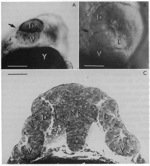
(A) Bifurcated eye in a living zebrafish embryo viewed with a dissecting microscope. The bifurcation (arrow) divides the eye into dorsal (D) and ventral (V) regions. In this case, pigment cells line the bifurcation. The embryo was treated 10.5 hr after fertilization with 1 μM retinoic acid for 1 hr and viewed 15 hr later. Y, yolk. (Bar = 140 μm.) (B) Bifurcated eye viewed by Nomarski optics in a living zebrafish embryo. Two retinas are readily visualized; one is located dorsally (D) to the bifurcation (arrow); the other is positioned ventrally (V). A lens (L) is associated with the ventral retina. The embryo in this case was treated at a late gastrula stage with 1 μM retinoic acid for 1 hr and raised, thereafter, in 0.2 mM phenylthiocarbamide to suppress pigment formation. The photomicrograph was taken 30 hr after fertilization. (Bar = 95 μm.) (C) Conventional light micrograph of a section through the head region of a zebrafish embryo. Dorsal (D) and ventral (V) retinas are present on both sides of the neural keel (NK). (Bar = 120 μm.) |

Light micrographs of eye sections from control (A) and retinoic acid-treated (B-D) zebraflish embryos. The treated embryos were exposed to 1 μM retinoic acid at the 2- to 3-somite stage for 2 hr. Drawings illustrating the key features of the photomicrographs are shown below. (A) Control showing a normally developing eyecup at the 17-somite stage. The thin (and difficult to see) optic lumen separates the pigment epithelial cell layer (PE) from the neural retinal epithelium (NR). Some mitotic figures are seen in the neural retina (arrow). (B) In the retinoic acid-treated embryo at the 17-somite stage, a dramatic expansion of the epithelial cell layer in the ventral portion of the eyecup is seen. Several mitotic figures are observed in this region (arrow). (C) By the 22-somite stage of development in a retinoic acid-treated embryo, two retinal epithelial cell layers can be distinguished. The optic lumen separates the dorsal (D) and ventral (V) retinas and the neural retinas from the pigment epithelial cells (PE). (D) By 24 hr after fertilization, the ventral retina (V) has assumed the same orientation as the dorsal retina (D). The calibration bar in A is 40 μm and applies to all parts of the figure. |

Ganglion cells and their axons stained with a monoclonal antibody in retinoic acid-treated animals. The embryos were treated with, 1 μM retinoic acid for 2 hr at the 2- to 3-somite stage and raised in 0.2 mM phenylthiocarbamide to suppress pigment formation. At 3.5 days of development, the embryo was fixed and processed for whole-mount staining. (A) Two fields (D and V) of ganglion cells can be seen in this retinoic acid-treated eye viewed with Nomarski optics from the side of the embryo. Bundles of axons project from each ganglion cell field and join to project centrally (arrow). (B) A view from the front of the head showing how the bundles of axons join as they exit the eye and innervate the contralateral tecta (T). (Bar = 170 μm.) |