
- Title
-
A pioneering growth cone in the embryonic zebrafish brain
- Authors
- Wilson, S.W. and Easter, S.S. Jr.
- Source
- Full text @ Proc. Natl. Acad. Sci. USA

Whole-mounted zebrafish brain showing labeled axons originating from the epiphysis at 24 hr. Rostral is to the right and dorsal is up. The epiphysial axons course ventrally in the DVDT to its intersection with the TPOC (dots), where they turn rostrally toward the POC. These are the only axons within the DVDT, whereas the TPOC contains others that run both rostrally toward the POC and caudally toward the midbrain (unlabeled in this preparation, but see ref. 17). E, epiphysis; POC, postoptic commissure; T, telencephalon; Te, tectum. (Bar = 50 μm.) |
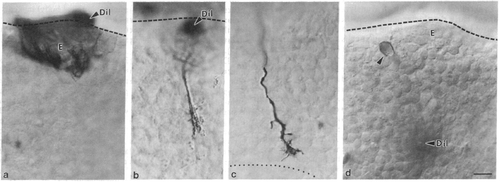
Micrographs from whole-mounted embryonic brains in which diI had been applied to the epiphysis (a-c) or to the DVDT (d). Rostral is to the right and dorsal is up. Dashed lines show the dorsal surface of the brain. (a) At 18 hr, cell bodies are labeled but none possesses axons or growth cones. (b) At 19.5 hr, a single pioneering growth cone is labeled approximately halfway down the DVDT. (c) At 21 hr, the pioneering growth cone is almost at the intersection with the TPOC (dotted line). (d) At 20 hr, a single neuron in the caudal epiphysis (arrowhead) is retrogradely labeled after diI application to the DVDT. E, epiphysis; DiI, application site. (Bar = 10 μm.) |
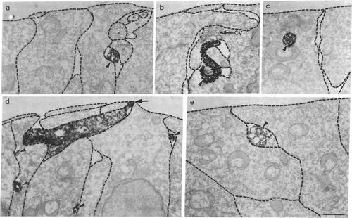
Electron micrographs of the pioneering axon and growth cone from preparations fixed at 19-20 hr. The basal lamina is toward the top of each panel. The dashed lines outline the boundaries of cells. (a) Transverse section through the labeled axon (arrowhead) near the epiphysis. Serial sections showed that the processes (asterisks) associated with it originated from nearby neuroepithelial cells. (b) Transverse section through the labeled axon (arrowhead) just proximal to the growth cone. A small side branch extends superficially and is apposed to a cluster of vesicles in the neuroepithelial cell that is enveloping the axon. Similar clusters were also seen in neuroepithelial cells untouched by the labeled structure, so they are probably not an artifact of the labeling. (c) Transverse section through a leading filopodium (arrowhead) that is inserted into a neuroepithelial cell. Penetrating filopodia, such as this, were not associated with coated vesicles, as has been described in insects (20), or with any other subcellular organelles. (d) Longitudinal section through a labeled growth cone that was advancing toward the right of the panel. The growth cone is in contact with the basal lamina (arrow). Filopodia (arrowheads) are between neuroepithelial cells, and some extend into the deeper nuclear layer of the neuroepithelium. The surface of the neuroepithelium is elevated above the growth cone, presumably a mechanical deformation. (e) Transverse section through the leading filopodium (arrowhead) of the growth cone. CV, cluster of vesicles. (Bar = 1 μm.) |