FIGURE SUMMARY
Title

Variants in NR6A1 cause a novel oculo vertebral renal syndrome

Authors
Neelathi, U.M., Ullah, E., George, A., Maftei, M.I., Boobalan, E., Sanchez-Mendoza, D., Adams, C., McGaughey, D., Sergeev, Y.V., Ai Rawi, R., Naik, A., Bender, C., Maumenee, I.H., Michaelides, M., Tan, T.G., Lin, S., Villasmil, R., Blain, D., Hufnagel, R.B., Arno, G., Young, R.M., Guan, B., Brooks, B.P.
Source
Full text @ Nat. Commun.

Phenotypes associated with variants in NR6A1.

a Pedigrees of three families (COL005; COL034; COL171) from the NEI cohort demonstrating coloboma with or without microphthalmia and cataract, missing vertebrae, and congenital renal anomalies. Inheritance is autosomal dominant with incomplete penetrance and variable expressivity. b Linear pigmentary disturbance representing a forme fruste of coloboma (arrow) in COL005.1 (right eye). c Larger chorioretinal coloboma in the left eye of COL005.1 demonstrating a retinal tear in the far periphery (arrowhead). d Iris coloboma of the left eye of COL005.10. e Microphthalmia of the left eye in COL034.1. f Retro-illumination image of the left eye of COL171.1 demonstrating iris coloboma and posterior subcapsular cataract (open arrow). g Spine x-ray of COL005.4 demonstrating 11 thoracic (normal 12) and 4 lumbar (normal 5) vertebrae. h Schematic of NR6A1 variants detected in the NEI and UK Genomics England cohorts. + individual with variant, − individual without variant. DNA binding domain (DBD) and putative nuclear receptor ligand binding domain (NR-LBD) are noted (Q15406; InterPro).

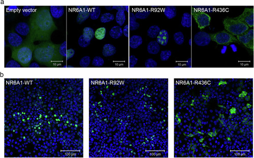
Subcellular localization of wild-type (WT) and mutant forms of NR6A1.

NR6A1 variant localization pattern was studied by overexpression in HEK293 cells and representative high magnification (63X) images are shown from three different trials (a) Scale bar = 10 µm. Low magnification images (b) scale bar 100 µm. The localization pattern for the WT and the two variant isoforms was observed to be consistent across three transfection experiments. (Cells counted: WT = 387, R92W = 350 and R436C = 217).

NR6A1 gene expression in ocular and body tissues.

a Comparative levels of NR6A1 from publicly available bulk human tissue RNA-sequencing (RNA-Seq) datasets accessed on the eyeIntegration website (https://eyeintegration.nei.nih.gov/). On average, expression is higher in embryonic and induced pluripotent stem cells (ESC, iPSC, respectively) than in adult ocular tissues. b Bulk RNA-Seq data in human retinal fetal tissue from two studies suggests NR6A1 expression is highest in early stages of development, including the window of optic fissure closure (lavender box). NR6A1 expression is plotted against the tissue age (days post conception, dpc). A linear regression analysis was added for each paper’s data from the 40 to 80 dpc and 80 to 160 dpc samples. c The density correlation plot (closer to −1 and 1 is more negatively or positively correlated, respectively) shows ten notable coloboma associated genes with highly ranked correlations with NR6A1 expression across eyeIntegration curated fetal retina and RPE tissues. This enrichment was not seen in adult tissues. d Among systemic tissues, NR6A1 is expressed most highly in bone marrow and testes. The boxplots display the median, 25th and 75th percentiles, and 1.5 * interquartile range (IQR). Any data outside the 1.5 * IQR are plotted. In panels a and d the number of samples is given above the boxplots.

Expression pattern of nr6a1a and nr6a1b paralogs in zebrafish.

nr6a1a is expressed ubiquitously at 11 hours post-fertilization (hpf) (a). By 16–19 hpf (b,c) expression is present in multiple structures including the somites (S), neural tube (NT) and notochord (N). At 24 hpf, expression remains in the NT but is decreased in the S and N. Expression in the lens (L) is first noted at 19 hpf and is particularly prominent by 24 hpf (dd”‘’). nr6a1b expression at 11 hpf (e) is in anterior trunk, localizing to neural tube and somites from 16 hpf (f) and 19 hpf (g). At 24hpf (hh”) it remains expressed in the neural tube and somites, with faint expression can be seen in the lens. All embryos are oriented in a lateral view, anterior to the left and dorsal up, except (d, d”, h’, and h”) shown in dorsal views. Scale bar = 100 µm. e-epiphysis, l-lens, p-pronephros, h- heart, n-notochord, s-somite, nt-neural tube, ad-anterior diencephalon, tg-tegmentum.

Rescue of nr6a1+nr6a1b zebrafish morphant phenotypes with wild-type and mutant human NR6A1 mRNA.

Controls (a, a’) have a straight body axis and the optic fissure (OF) is closed. The nr6a1+nr6a1b morphants that have a mild phenotype (b, b’) have close to a normal body with microphthalmia and heart edema; a moderate phenotype (c, c’) with a slightly bent body axis with smaller eyes, coloboma and a severe heart edema; and severe morphants (d, d’) have a curved body axis with smaller eyes, coloboma and heart edema. The morphant phenotype was rescued when the morpholinos were co-injected along with the human-NR6A1-wild-type mRNA. However, there was no significant rescue in the morphant phenotype when the morpholinos were injected with either R92W or R436C human disease-causing variants (e). Morpholinos were injected at 0.75 ng each (1.5 ng total). Embryos were imaged at 72 hpf. Scale bar = 100 µm. Statistical significance was calculated using Chi-square test and Fisher’s test. P value of nr6a1-TB-MO(a + b) V hWT-NR6A1 is <0.0001 and between nr6a1-TB-MO(a + b) V hR92W and hR436C are 0.0266 and 0.5169 respectively. Source data file has been provided for (e).

nr6a1(a + b) zebrafish morphants have kidney and vertebrae defects.

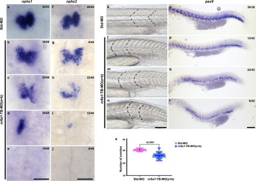
At 48 hpf, control embryos demonstrate bilateral expression of nphs1 (nephrin) and nphs2 (podocin) (a, f), markers of kidneys. Knockdown of nr6a1(a + b) resulted in a range of abnormal expression patterns including mildly reduced expression (b, g), moderately reduced expression (c, h), unilateral or midline expression (d, i) or absent expression (e, j) of nphs1 and nphs2. Somites (the early precursors of vertebrae) have a sharp, chevron shape with an approximately 90-degree angle in control zebrafish at 24 hpf (k). Knockdown of nr6a1a and nr6a1b resulted in a range of abnormal expression patterns including chevron shapes with obtuse angles (l), as well as mildly rounded (m) or flattened (n) somites. The sclerotome marker pax9 is expressed in a continuous angular pattern in the ventromedial portion of somites along the length of 24 hpf control embryos (o). Double morphant embryos exhibit a spectrum of abnormal patterns of pax9 expression including reduced levels of expression (p), patchy expression extending beyond the caudal tip of the yolk (q) and patchy expression ending near the caudal end of the yolk sac (r). nr6a1(a + b) morphants have reduced number of somites (s), statistical significance is calculated using two tailed t-test. Std-MO n = 20 and nr6a1-TB-MO(a + b) n = 50. Data is represented as difference between means of nr6a1-TB(a + b)–Std-MO ± SEM (−4.490 ± 0.5629). P value is <0.0001. 0.75 ng of each paralogue is used for knock-down. Numbers of each representative pattern are given in each panel. Scale bar = 100 µm. Source data file has been provided for (s).

EXPRESSION / LABELING:
Genes:
Fish:
Knockdown Reagents:
Anatomical Terms:
Stage Range: Prim-5 to Long-pec
PHENOTYPE:
Fish:
Knockdown Reagents:
Observed In:
Stage Range: Prim-5 to Long-pec
Acknowledgments
This image is the copyrighted work of the attributed author or publisher, and ZFIN has permission only to display this image to its users. Additional permissions should be obtained from the applicable author or publisher of the image. Full text @ Nat. Commun.