FIGURE SUMMARY
Title

A Novel De Novo Missense Variant in Netrin-1 (NTN1) Associated With Chorioretinal Coloboma, Sensorineural Hearing Loss and Polydactyly

Authors
Toms, M., Heppell, C., Owen, N., Malka, S., Moosajee, M., Genomics England Research Consortium
Source
Full text @ Clin. Genet.

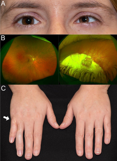
Clinical features of the patient with a heterozygous missense variant c.1483T>A p.(Tyr495Asn) in NTN1. (A) Facial photo showing unilateral left microphthalmia and microcornea. (B) Widefield colour fundus imaging showing extensive left chorioretinal coloboma involving the optic disc and macula, right fundus is normal. (C) Clinical photograph of both hands with white arrow pointing to right hand indicating site of extra digit (polydactyly), which was surgically removed from between ring and little finger.

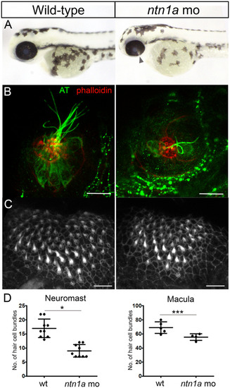
Ocular coloboma and sensory hair cell defects in ntn1a morphant zebrafish. (A) Fusion of the optic fissure was not complete in ntn1a morphant embryos at 56 h post‐fertilisation. The arrow indicates the ocular coloboma. (B) The lateral line neuromasts were visualised in control and ntn1a larvae at 5 days post‐fertilisation (dpf). Anti‐acetylated tubulin (AT, green) detected the hair cell bodies and kinocilia and Alexa Fluor 647 Phalloidin (red) stained the stereociliary hair bundles. The ntn1a neuromasts lacked hair cell bodies, hair bundles and kinocilia. (C) The stereociliary hair bundles of the anterior macula in the inner ear were visualised used Alexa Fluor 647 Phalloidin (white). The hair bundle morphology appeared to be normal in ntn1a at 5 dpf, but the hair bundles were reduced in number. (D) The bar charts show that the numbers of hair bundles per neuromast (n = 9, *p < 0.05) and per anterior macula (n = 5, ***p < 0.001) were significantly reduced in the ntn1a morphant compared with control larvae at 5 dpf. Scale bars = 10 μm.

Acknowledgments
This image is the copyrighted work of the attributed author or publisher, and ZFIN has permission only to display this image to its users. Additional permissions should be obtained from the applicable author or publisher of the image. Full text @ Clin. Genet.