
- Title
-
Exposure to valproic acid (VPA) reproduces hdac1 loss of function phenotypes in zebrafish
- Authors
- Jones, A.A., Willoner, T., Mishoe Hernandez, L., DeLaurier, A.
- Source
- Full text @ MicroPubl Biol
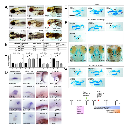
Exposure to VPA produces defects in heart, eye, pectoral fin, and craniofacial development consistent with hdac1 loss of function mutants and is associated with changes to tbx5 gene expression: (A) Phenotypes associated with VPA treatment (0.5 and 1.0mM) compared with controls and hdac1 mutant larvae (top row, lateral views: heart indicated by black arrow; middle row lateral views: eye indicated by red arrow; bottom row dorsal views: pectoral fin indicated by green arrow). Bar = 200 microns. (B) Frequency of phenotypes associated with specific VPA exposures (0.5 and 1.0mM) in 6 dpf larvae. (C) RT-qPCR results showing relative expression changes in tbx5a and tbx5b in VPA (0, 0.5, and 1.0mM) treated embryos compared to controls (at 30 and 54 hpf). Expression of tbx5 genes was standardized against expression of the housekeeping gene eif1a and levels of expression are relative to controls. Average of 5 biological replicates and 3 technical replicates per group. Data was analyzed using a Kruskal-Wallis test to examine the effects of VPA concentration on gene expression levels. A Dunn’s multiple comparison test was used to determine statistical differences between VPA concentrations at each stage, (* p < 0.05, n.s. = not significant). Lines indicate significance between groups. (D) mRNA in situ hybridization showing expression of tbx5a, tbx5b, and fgf24 in VPA treated embryos (0.5 and 1.0 mM) and controls (first and second row left columns: pectoral fin; third and fourth row left columns eyes; first row right: heart region; second through fourth row right columns: pectoral fin). Dotted lines show lateral margin of outgrowth of pectoral fin buds in VPA-treated embryos. In each experiment expression was assessed in at least 10 stained embryos and representative images are shown. (E) Skeletal preparations (lateral view) of control larvae at 72 (n = 34), 84 (n = 33), and 96 (n = 22) hpf (mk = Meckel’s; pq = palatoquadrate; hs = hyosymplectic; ch = ceratohyal; op = opercle; bsr = branchiostegal ray). Cartilage is stained blue (Alcian Blue) and bone is stained red (op and bsr, Alizarin Red). Bar = 200 microns. (F-G) Skeletal preparations of VPA-treated (0.5 mM) and hdac1 mutant larvae where (F) VPA was added to embryos at bud stage (10 hpf) and removed at 24 (n = 49), 36 (n = 89), 48 (n = 8), and 60 (n = 59) hpf (lateral and ventral views) compared to the hdac1 mutant; (G) VPA was added at 24 (n = 32), 36 (n = 46), 48 (n = 42), 60 (n= 46), and 72 (n = 44) hpf and removed at 96 hpf (lateral views). All VPA-treated larvae were fixed at 96 hpf, and the hdac1 mutant larvae were fixed at 5 dpf. Cartilage is stained blue (Alcian Blue) and bone is stained red (op and bsr, Alizarin Red). Arrows are pointing to pharyngeal cartilages in ventral views. Bar = 200 microns. (H) Timeline showing events associated with pharyngeal skeleton development and hypothesized role of Hdac1. dpf = days post-fertilization hpf = hours post-fertilization |