FIGURE SUMMARY
Title

The prolyl isomerase Pin1 stabilizes NeuroD during differentiation of mechanoreceptors

Authors
Zhao, L., Fong, S.H., Yang, Q., Jiang, Y.J., Korzh, V., Liou, Y.C.
Source
Full text @ Front Cell Dev Biol

Sequence analysis of zebrafish Pin1. (A) The full-length cDNA sequence of zebrafish pin1. Red letters indicate 5′-UTR region; blue letters indicate 3′-UTR region; and black letters indicate the open reading frame. (B) Amino acid sequence alignment of Pin1 in Drosophila (dodo), budding yeast (Ess1), Xenopus (xPin1), zebrafish (zPin1), mouse (mPin1) and human (hPin1). Conserved residues in all species are highlighted in red. Residues with at least 50% identity across species are highlighted in blue. The numbers refer to amino acid positions of zebrafish Pin1.

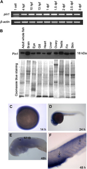
The spatial and temporal expression patterns of zebrafish Pin1. (A) Semiquantitative RT-PCR analysis of pin1 expression in different stages of zebrafish development. β-actin served as an internal control. (B) Western blot analysis of Pin1 expression in adult zebrafish tissues. Coomassie Blue staining served as internal control. Each lane contains 30 µg of total protein. (C–F) WISH analysis of pin1 expression in 14 hpf embryo (C), 24 hpf (D) and 48 hpf embryo (E,F). Arrow in (E) indicate midbrain-hindbrain boundary (MHB). Arrows in (F) mark the neuromasts staining in the PLL system.

Zebrafish embryonic developmental delay in pin1 morphants. (A) The design scheme of pin1 MO1 and MO2. ORF: Open reading frame. (B) Pin1 protein levels in zebrafish embryos injected with standard control MO and pin1 MO at the concentration indicated. β-tubulin was used as loading control. (C–J) Depletion of Pin1 results in embryonic developmental delay.

Specific PLL neuromasts hair cells defects in pin1 morphants. (A–F) Co-injection of pin1 MO with p53 MO results in slight developmental delay and defective neuromasts hair cells formation in zebrafish embryos. Arrows in (C) indicate posterior neuromasts. (G–I) Analysis of neuromasts cells in pin1 MO/p53 MO injected ET4 embryos. Arrows in (G, H) indicate neuromasts hair cells along the PLL. (J–L) Analysis of mantle cells in pin1 MO/p53 MO injected ET20 embryos. Arrowheads in (J–L) indicate neuromasts mantle cells. (M–P) Analysis of hair cells and mantle cells in 48 hpf and 72 hpf double transgenic (ET4x20) embryos.

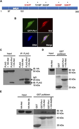
Zebrafish Pin1 interacts with Nrd via pSer/Thr-Pro motifs. (A) A schematic illustration of the potential Pin1-binding sites in Nrd. (B) Zebrafish Pin1 and Nrd co-localizes in the SH-SY5Y cell nucleus. DNA (blue), zebrafish Pin1 (green), Nrd (red). (C,D) Zebrafish Pin1 interacts with Nrd in vivo and in vitro.(E) The interaction of HA-Nrd5A mutant and zebrafish Pin1 is compromised.

Pin1 regulates Nrd stability. Protein stability assay of HA-Nrd in HEK 293T cells stably expressing control siRNA and Pin1 siRNA. (A) HA-Nrd degraded at a higher rate in Pin1 knock-down background in HEK 293T cell line. (B) HA-Nrd5A levels remained stable in Pin1 knock-down background in HEK 293T cells. (C) Overexpression of FLAG-tagged zebrafish Pin1 enhanced Nrd stability. α-tubulin was used as an internal control. (D) Quantification of HA-Nrd levels in (A,C) normalized to α-tubulin levels. (E) Quantification of HA-Nrd levels in (B) normalized to α-tubulin levels. Error bars represent ±SEM.

Acknowledgments
This image is the copyrighted work of the attributed author or publisher, and ZFIN has permission only to display this image to its users. Additional permissions should be obtained from the applicable author or publisher of the image. Full text @ Front Cell Dev Biol