
- Title
-
Zebrafish thrombosis models according to the location of thrombus formation
- Authors
- Hwang, J., Koun, S., Ha, Y., Jung, J.M.
- Source
- Full text @ Ann Transl Med

In vivo images at pre-and post-photoactivation. (A) In vivo cerebral angiography of vascular endothelial cells in transgenic zebrafish larvae [Tg (flk:gfp)] (GFP). (B) Evaluation of blood flow using the 560 nm laser fluorescence microscope (rhodamine). (C) Evaluation of blood flow in the endothelial lining by merging GFP and rhodamine. Blood flow in intracerebral vessels disappeared, and endothelial linings’ fluorescence intensity slightly decreased (D-F). GFP, green fluorescent protein. |

Real-time imaging of thrombosis in the vasculature (in vivo angiographies and blood flow chart per cross-sectional area of total vasculatures). Compared with the pre-photoactivation state (A,C), no thrombus or change of blood flow was observed at the post-activation state in the control group after photocoagulation (B), while thrombus formation and decreased blood flow were observed after photocoagulation in the Rose Bengal group (D). |
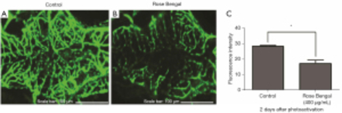
Analysis of vascular endothelial cell apoptosis at pre-and post-photoactivation (after 2 days). (A) In vivo angiography of the control group, (B) in vivo angiography of photosensitizer injection group, (C) fluorescence intensity: control: 27.83±0.56 (mean ± standard deviation) photocoagulation (Rose Bengal + rhodamine): 17.28±2.08 (measured by Image J 1.52a software). *, P<0.01. |

Real-time imaging of the thrombolytic activity of tPA. (A) Thrombus formation after photocoagulation, (B) thrombolytic activity of tPA, (C) comparison of thrombus area using Image J 1.52a (Mann Whitney test, P<0.05). Rose Bengal + tPA (n=4) group shows a decreased thrombus area (49.78%±7.090%, mean ± standard deviation). *, P<0.05. tPA, tissue plasminogen activator. |

Quantitative assay of intracardiac thrombosis. (A) Control group, (B) Rose Bengal group, (C) Rose Bengal + tPA group, (D) comparison of intracardiac thromboses, red circles indicate thrombus in the zebrafish heart shown by O-dianisidine staining of the red blood cells. *, P<0.05; **, P<0.01. tPA, tissue plasminogen activator. |