FIGURE SUMMARY
Title

A median fin derived from the lateral plate mesoderm and the origin of paired fins

Authors
Tzung, K.W., Lalonde, R.L., Prummel, K.D., Mahabaleshwar, H., Moran, H.R., Stundl, J., Cass, A.N., Le, Y., Lea, R., Dorey, K., Tomecka, M.J., Zhang, C., Brombacher, E.C., White, W.T., Roehl, H.H., Tulenko, F.J., Winkler, C., Currie, P.D., Amaya, E., Davis, M.C., Bronner, M.E., Mosimann, C., Carney, T.J.
Source
Full text @ Nature

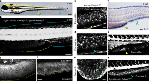
A non-PM-derived median fin fold.

a, Larval 4 dpf zebrafish possess a median PAFF (yellow arrowhead) in addition to the caudal median fin fold (cyan arrowhead). b, Confocal image of a 3 dpf Tg(tbx16l:GAL4-VP16); Tg(UAS:Kaede) embryo with PM labelled by Kaede showing PM-derived mesenchyme in the caudal median fin fold (cyan outline) but not the PAFF (yellow outline). ce, Confocal images of pre-anal (c), ventral caudal (d) and pectoral (e) fins of the ET37 Enhancer Trap transgenic line indicating that PAFF contains morphologically comparable mesenchyme (indicated by arrowheads) to other larval fin folds. f, In situ hybridization of the fin mesenchyme marker fbln1 in both PAFF (yellow arrowhead) and caudal fin fold (cyan arrowhead) at 3 dpf. g,h, DsRed expression in mesenchyme of both pre-anal (g) and caudal (h) fin folds of the 5 dpf Tg(-5.2lyve1b:DsRed) transgenic line. i,j, Immunostaining for collagen II in 8 dpf frf mutants (j) shows loss of fibril organization compared with WT (i). Scale bars, 200 µm (a); 100 µm (b); 20 µm (c,e); 50 µm (f,g,j).

The PAFF is an LPM-derived median fin fold.

a,b, Confocal images of Tg(hand2:EGFP) embryos at 2 dpf (a) and 3 dpf (b) showing eGFP labelling of the mesenchyme of the pectoral (green outline and arrowheads) and PAFFs (yellow outline and arrowheads, and magnified in inset) but not the caudal fin fold (cyan outline). c,d, In situ hybridization of hand2 at 3 dpf shows fin expression of hand2 only in the PAFF (yellow arrowheads (c), and higher magnification with Nomarski optics indicates expression in the mesenchyme (d). e, Schematic of the LPM lineage tracing transgenes. f, Lineage tracing of LPM using transgenics depicted in (e) following 4-OHT treatment and heat shock before imaging shows that PAFF mesenchyme is derived from the LPM (yellow arrowhead and magnified in inset). g,h, Ventral (g) and lateral (h) confocal images of the drl:H2B-Dendra2 transgenic line at the 10-somite stage (10 ss) (g) and 48 hpf (h) following ultra-violet laser photoconversion in the region of the LPM outlined in (g). h, Photoconverted PAFF mesenchyme is indicated by yellow arrowheads. Scale bars, 100 µm (a,c,f); 50 µm (a (inset),f (inset),g,h); 200 µm (b); 20 µm (d).

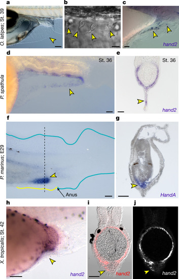
PAFF mesenchyme expression of hand2 is conserved across vertebrates.

a,b, Nomarski images of 9 dpf medaka showing PAFF (a) with dispersed mesenchymal cells (yellow arrowheads) (b). c, In situ hybridization of medaka at stage 39 showing hand2 expression in pre-anal fin mesenchyme (yellow arrowheads). d,e, In situ hybridization of hand2 in stage 36 paddlefish embryos shown laterally (d) or in transverse section (e). hand2-positive PAFF fin mesenchyme is indicated by yellow arrowheads. f,g, HandA in situ hybridization of stage E29 lamprey (P. marinus) embryos shown laterally (f) or in transverse section (g). Lamprey show strong expression of HandA in a fin anterior to the anus (yellow arrowhead in f) corresponding to cells in the interior of the fin (g). The section location of (g) is indicated by the dashed line in (f). hj, Chromogenic (h) or fluorescent (i,j) in situ hybridization of hand2 in stage 42 X. tropicalis embryos shown laterally (h) or in transverse section (i,j). Fluorescent image (j) is overlayed on the Nomarski image (i). The small PAFF in Xenopus contains sparse hand2-positive fin mesenchyme (yellow arrowheads). St., stage. Scale bars, 100 µm (a,d,h); 10 µm (b); 20 µm (c); 50 µm (eg,i).

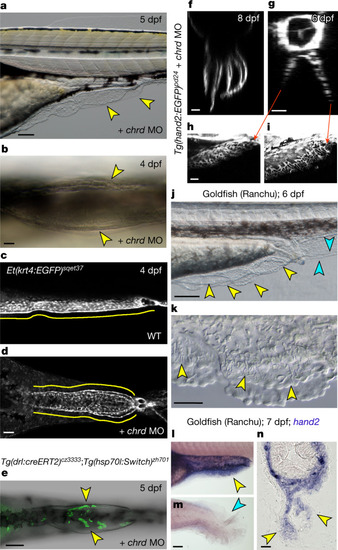
Duplication of the PAFF into paired fin folds.

a,b, Lateral (a) and ventral (b) Nomarski images of PAFFs of 5 dpf (a) and 4 dpf (b) embryos injected with MO targeting chordin, resulting in duplication of PAFF (yellow arrowheads). c,d, Confocal micrographs, imaged ventrally, of PAFFs in 4 dpf ET37 transgenic larvae uninjected (c) or injected with a chrd MO (d). PAFFs are indicated with yellow lines. d, Note the duplicated anal openings following chrd MO injection. e, Ventral confocal image of chrd MO duplicated PAFFs of the transgenic line shown above. Hydroxytamoxifen treatment at the 12-somite stage and heat shock before imaging show that mesenchyme of duplicated PAFFs is derived from the LPM (yellow arrowheads). fi, Light-sheet (f) and confocal (gi) images of Tg(hand2:EGFP) larvae at 8 dpf (f) and 6 dpf (gi). Orthogonal display through the xz plane (f,g) shows that multiple PAFFs can form and that duplicated PAFFs contain eGFP-positive mesenchyme (gi). Lateral views of left (h) and right (i) duplicated PAFFs of sample in g are given. j,k, Lateral low- (j) and high-power (k) Nomarski images of the PAFF of the Ranchu goldfish at 6 dpf. The multiple PAFFs (j) and individual mesenchyme cells (k) are indicated by yellow arrowheads. Duplicated caudal fin folds are indicated by cyan arrowheads (j). ln, Lateral (l,m) and transverse (n) views of pre-anal (l,n) and caudal (m) fin folds of 7 dpf Ranchu larvae stained by in situ hybridization for hand2, where hand2-positive PAFF mesenchyme is indicated by yellow arrowheads. Absence of hand2 in the caudal fin fold is indicated by the cyan arrowhead (m). Scale bars, 100 µm (a,e,m); 20 µm (b,fh,n); 50 µm (d,k); 200 µm (j).

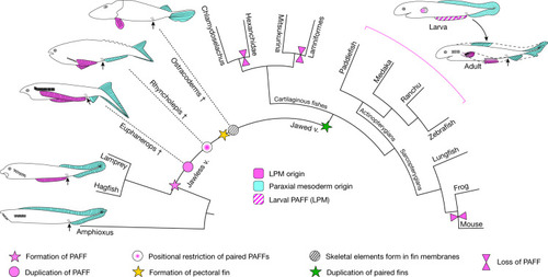
Hypothesis of the elaboration of the PAFF to paired fins.p>Simplified evolutionary scenario of vertebrates showing the presence of a PAFF and subsequent modifications leading to paired fins. Dashed lines and dagger symbols indicate extinct lineages, and solid lines indicate extant lineages. PM-derived fins and fin folds are in cyan, while LPM-derived fins are in pink. Larval PAFF is hatched. Black arrows indicate the position of the anus.

Acknowledgments
This image is the copyrighted work of the attributed author or publisher, and ZFIN has permission only to display this image to its users. Additional permissions should be obtained from the applicable author or publisher of the image. Full text @ Nature