IMAGE

Fig. 3

ID
ZDB-IMAGE-230617-73
Source
Figures for Tzung et al., 2023
Image
Figure Caption

Fig. 3 
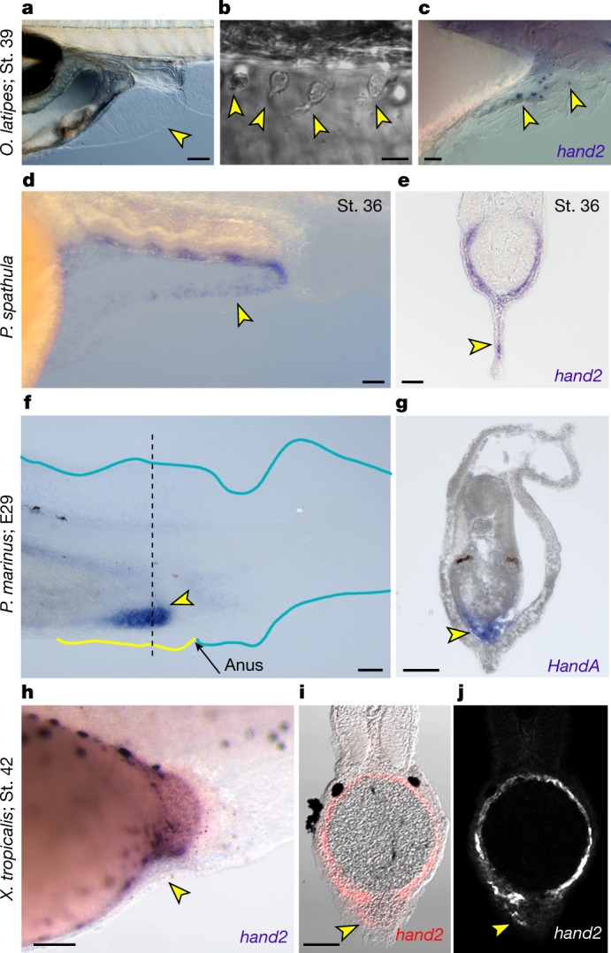
PAFF mesenchyme expression of hand2 is conserved across vertebrates.

a,b, Nomarski images of 9 dpf medaka showing PAFF (a) with dispersed mesenchymal cells (yellow arrowheads) (b). c, In situ hybridization of medaka at stage 39 showing hand2 expression in pre-anal fin mesenchyme (yellow arrowheads). d,e, In situ hybridization of hand2 in stage 36 paddlefish embryos shown laterally (d) or in transverse section (e). hand2-positive PAFF fin mesenchyme is indicated by yellow arrowheads. f,g, HandA in situ hybridization of stage E29 lamprey (P. marinus) embryos shown laterally (f) or in transverse section (g). Lamprey show strong expression of HandA in a fin anterior to the anus (yellow arrowhead in f) corresponding to cells in the interior of the fin (g). The section location of (g) is indicated by the dashed line in (f). hj, Chromogenic (h) or fluorescent (i,j) in situ hybridization of hand2 in stage 42 X. tropicalis embryos shown laterally (h) or in transverse section (i,j). Fluorescent image (j) is overlayed on the Nomarski image (i). The small PAFF in Xenopus contains sparse hand2-positive fin mesenchyme (yellow arrowheads). St., stage. Scale bars, 100 µm (a,d,h); 10 µm (b); 20 µm (c); 50 µm (eg,i).

Acknowledgments
This image is the copyrighted work of the attributed author or publisher, and ZFIN has permission only to display this image to its users. Additional permissions should be obtained from the applicable author or publisher of the image. Full text @ Nature