
- Title
-
Early development of respiratory motor circuits in larval zebrafish (Danio rerio)
- Authors
- McArthur, K.L., Tovar, V.M., Griffin-Baldwin, E., Tovar, B.D., Astad, E.K.
- Source
- Full text @ J. Comp. Neurol.

High-speed behavioral imaging demonstrates that larval zebrafish can produce bouts of patterned operculum movements at 3 days postfertilization (dpf). (a) At 3 dpf, some fish (n = 2 of 13) exhibited irregular operculum movements of varying amplitude and frequency, without a clear pattern. (b) Most fish at 3 dpf (n = 11 of 13) exhibited bouts of rhythmic operculum movement, followed by a large burst of operculum movement associated with attempted whole-body movement (indicated by arrows), followed by a pause before rhythmic movement resumed. In both panels (a) and (b), a microscopic image of the operculum is shown in the upper left, and a representative trace of dorsoventral operculum position over time is shown in the upper right. The location of the landmark used to track movements in this subject is indicated by an asterisk. A histogram (bottom) shows the distribution of inter-movement intervals for this subject across trials (excluding intervals during and immediately following attempted whole-body movements). The histogram has been divided into smaller (<5 s, left) and larger (>5 s, right) intervals. |

Operculum behavior becomes more consistently patterned at 4 and 5 days postfertilization (dpf). At both (a) 4 dpf (n = 15) and (b) 5 dpf (n = 15), all subjects produced bouts of rhythmic operculum movement, followed by a large burst of operculum movement associated with attempted whole-body movement (indicated by arrows), followed by a pause before rhythmic movement resumed. Note that rhythmic movements could occur as couplets, as shown for the example in panel (b). In both panels (a) and (b), a microscopic image of the operculum is shown in the upper left, and a representative trace of dorsoventral operculum position over time is shown in the upper right. The location of the landmark used to track movements in this subject is indicated by an asterisk. A histogram (bottom) shows the distribution of inter-movement intervals for this subject across trials (excluding intervals during and immediately following attempted whole-body movements). The histogram has been divided into smaller (<5 s, left) and larger (>5 s, right) intervals. |

Bouts of patterned operculum movements at 3 days postfertilization (dpf) have similar temporal properties overall to those observed at 4 and 5 dpf. Histograms show the distribution of operculum inter-movement intervals across subjects at (a) 3 dpf (n = 13; 4842 total intervals), (b) 4 dpf (n = 15; 13,657 total intervals), and (c) 5 dpf (n = 15; 11,637 total intervals). The number of total intervals falling within a particular range (in seconds) is shown as a percentage of the total number of intervals in that age group. Intervals during and immediately following attempted whole-body movements were excluded from this analysis, to focus on the rhythmic component of the behavior. Fish produced short-interval operculum movements (<1 s) at all three ages, though fish exhibited many more long (>5 s) intervals at 3 dpf than at 4 or 5 dpf. |
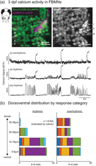
Calcium imaging demonstrates that facial branchiomotor neurons (FBMNs) exhibit both rhythmic and nonrhythmic activity at 3 days postfertilization (dpf), and reveals dorsoventral topography by activity pattern. (a) At 3 dpf, some FBMNs (i) exhibit only large, infrequent (“nonrhythmic”) calcium transients associated with attempted whole-body movements (indicated by arrows), while other FBMNs (ii and iii) exhibit bouts of additional burst activity (“rhythmic”). Sampling interval = 295 ms. (b) At 3 dpf, rhythmic FBMNs are located almost exclusively in the ventral half of the facial motor nucleus (0–19 μm from the ventral-most FBMN, for each fish), while nonrhythmic FBMNs are found throughout its dorsoventral extent. Response category (rhythmic vs. nonrhythmic) is a significant predictor of dorsoventral position (n = 6 fish; mixed effects linear model: p = .0035). Bars of the same color indicate FBMNs imaged in the same fish. Dorsoventral position is measured with respect to the approximate plane of the bilateral facial motor nerve within the hindbrain. |

Operculum movements are coordinated with pectoral fin movements at both 3 and 5 days postfertilization (dpf). (a) At 3 dpf, all subjects (n = 9) produced bouts of high-frequency (10–35 Hz) pectoral fin movements, typically accompanied by one or more operculum movements. (b) At 5 dpf, all subjects (n = 8) produced shorter bouts of high-frequency (10–50 Hz) pectoral fin movements, almost always accompanied by one or more operculum movements. Operculum movements rarely occurred in the absence of pectoral fin movement, at either age. In both panels (a) and (b), a lateral view microscopic image of the subject is shown in the upper left, accompanied by a line drawing that highlights key anatomical features used to track fin and operculum movements. The plots underneath the images show the peak displacement times for the operculum and pectoral fin during a 30-s period, represented as vertical lines. |

Operculum–fin coordination becomes tighter and less variable between 3 and 5 days postfertilization (dpf). Histograms show the distribution of delays between pectoral fin and operculum movements at (a) 3 dpf (n = 9; 325 fin–operculum delays shown, from 470 total fin bouts—145/470 fin bouts occurred without operculum movement) and (b) 5 dpf (n = 8; 1107 total fin–operculum delays shown, from 1108 total fin bouts—1/1108 fin bouts occurred without operculum movement). Fin-to-operculum delay was calculated as the difference in the peak displacement time of the first pectoral fin and operculum movements, within a single pectoral fin bout. A positive value indicates that the fin reached peak displacement before the operculum, and a negative value indicates that the operculum reached peak displacement before the fin. Note that most delay values are positive, indicating that the fin typically leads the operculum. This delay was longer overall at 3 dpf (median delay = 0.17 s) than 5 dpf (median delay = 0.03 s), confirmed by statistical comparison (mixed effects linear model: p < .0001). |