
- Title
-
X-linked variations in SHROOM4 are implicated in congenital anomalies of the urinary tract and the anorectal, cardiovascular and central nervous systems
- Authors
- Kolvenbach, C.M., Felger, T., Schierbaum, L., Thiffault, I., Pastinen, T., Szczepańska, M., Zaniew, M., Adamczyk, P., Bayat, A., Yilmaz, Ö., Lindenberg, T.T., Thiele, H., Hildebrandt, F., Hinderhofer, K., Moog, U., Hilger, A.C., Sullivan, B., Bartik, L., Gnyś, P., Grote, P., Odermatt, B., Reutter, H.M., Dworschak, G.C.
- Source
- Full text @ J. Med. Genet.
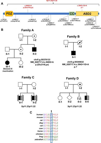
Exome sequencing and copy number variation analyses in families with congenital malformations identify variants in SHROOM4. (A) Protein domain structures are depicted (https://smart.embl.de/). The position of newly identified single-nucleotide variants of families A and B and the partial deletion of SHROOM4 in families C and D are annotated in red. Previously reported single-nucleotide variants are shown in black. (B) Pedigree with two affected individuals in family A . III-1 presented with several congenital malformations with her maternally derived X-chromosome being activated in 84% of all lymphocytes. Pedigrees of families C and D that bear microdeletions including CLCN5 and SHROOM4 are depicted. Healthy carriers of variants are highlighted with a dot. Symbols representing affected individuals are shaded with different fill for differentiating clinical conditions. Black boxes depict the congenital malformation phenotype. Striped boxes illustrate Dent’s disease. Brackets denote adoption. Triangle denotes miscarriage. (C) Amino acid sequence conservation among species of p.Glu314Lys that was altered in SHROOM4 in family A. CC, coiled coil; wk, gestational age in weeks. |
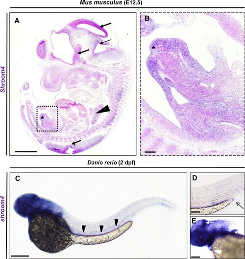
Shroom4/shroom4 is expressed during early mouse and zebrafish development. (A,B) ISH with Shroom4 probe on a sagittal section of a representative E12.5 (TS21) Mus musculus. Shroom4 expression is visible (in blue) in the head and neuronal tissues (black arrows), Vertebrae (black arrowhead), and genital tubercle (asterisks). Magnification (square in A) highlights the expression in the developing genitourinary tract (B). (C) Whole-mount ISH with an anti-shroom4 probe shows the expression of shroom4 RNA (in blue) at 2 days post fertilisation in the head, brain, eyes, fins, heart, the intestine (black arrowheads) and cloacal region in Danio rerio. Sense controls did not show a staining (not shown). (D) The cloaca is highlighted by an arrow in the enlargement. (E) The expression of shroom4 is highlighted by a white arrow in the brain and by a white arrowhead in the heart in the enlargement. Positive expression in the pectoral fins is marked by the white asterisk. Scale bars represent 1 mm (A), 10 µm (B) and 100 µm (C). ISH. In situ hybridisation. |

Knockdown of Shroom4 leads to glomerular cysts, eye abnormalities and PE. (A) Western blot analysis demonstrates a protein decrease in shroom4 MO-injected zfl for Shroom4 (184 kDa, F1Q6C1), derived from transcript shroom4-201, in comparison with Ctrl MO-injected zfl. The protein product for transcript shroom4-202 is too small to be detected by Western blot analysis (11 kDa, B3DGK9). β-Actin serves as Ctrl and shows an equal amount of protein in both samples. (B) Quantification of survival (n=5); zfl injected with shroom4 MO show a significant reduction of survival rate at 5 dpf compared with Ctrl MO. Survival of shroom4 MO is significantly rescued by coinjection of human WT SHROOM4 RNA. No alterations in survival have been observed by solely injecting WT SHROOM4 RNA. (C,D) Quantification of PE (n=3) shows significant occurrence in shroom4 morphants, a phenotype, which could be rescued by co-njection of WT SHROOM4 RNA. Black arrowhead highlights PE observed in a zfl injected with shroom4 MO at 4 dpf. (E,F) Eye-to-head ratio of injected zfl at 4 dpf (n=4). Measurement of the eye (red line) and head (black line, distance between the snout to the otic vesicle) was performed as visualised (F). Injection of shroom4 MO significantly reduced eye-to-head ratio, while WT mRNA co-injection in shroom4 MO-injected zfl significantly rescues the phenotypical effect. (G,H) Zfl injected with shroom4 MO develop glomerular cysts (white arrowheads in H) and dilatation of the pronephric ducts (white asterisks in H) that could not be rescued by human WT RNA (n=4). Images from in vivo observation through fluorescence microscopy (dorsal view) in Tg(wt1b:GFP) were taken at 2 dpf. Scale bars represent 100 µm. Ctrl, control; MO, morpholino oligonucleotide; ns, nonsignificant; PE, pericardial effusion; wt, wild type; zfl, zebrafish larvae. p <0.05 (*), p <0.01 (**), p <0.001 (***) |
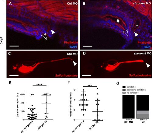
Phalloidin stainings and SR101 excretion assay show cloacal obstruction in shroom4 morphants. (A,B) Cloacal region is depicted at 5 dpf. White arrows indicate the hindgut; the white arrowheads show the distal pronephric ducts. With shroom4 MO-injected zfl display a dilated hindgut and distal pronephric morphology compared with Ctrls. (C,D) KD with shroom4 MO causes an abnormal cloacal excretion of SR101 compared with Ctrl MO-injected zfl at 5 dpf elucidated by white arrowheads (C,D). (E–G) shroom4 morphants show significantly longer time to excretion (E), significantly reduced number of excretions over time (F), and oscillating or missing peristalsis compared with Ctrls (G) (n=3). Scale bars represent 30 µm (A,B) and 100 µm (C,D). Ctrl, control; MO, morpholino oligonucleotide; SR101, sulforhodamine 101; zfl, zebrafish larvae. |