
- Title
-
Deciphering sex-specific miRNAs as heat-recorders in zebrafish
- Authors
- van Gelderen, T.A., Montfort, J., Álvarez-Dios, J.A., Thermes, V., Piferrer, F., Bobe, J., Ribas, L.
- Source
- Full text @ Sci. Rep.
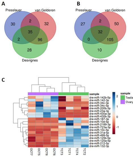
The number of miRNAs that were significantly expressed (average normalized reads > 100) of two AB strains (current data and Desvignes et al. |
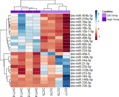
Heatmap of 23 differentially expressed (DE) miRNAs in mature ovaries after exposing zebrafish to high temperature during sex differentiation. The color scale ranges from blue to red, where blue shows relative overexpression and red is relative under expression. Both miRNAs and samples were grouped by hierarchical clustering. |
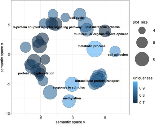
Visual representation of Gene Ontology (GO) terms related to Biological process obtained from predicted target genes of differentially expressed miRNAs in ovary. The most frequent terms were regulation of transcription, transport and phosphorylation. Color intensity represents the uniqueness of the GO term. Plot_size shows the frequency of the GO term in the UniProt database. The GO terms with a dispensability of < 0.25 are annotated in the plot. |
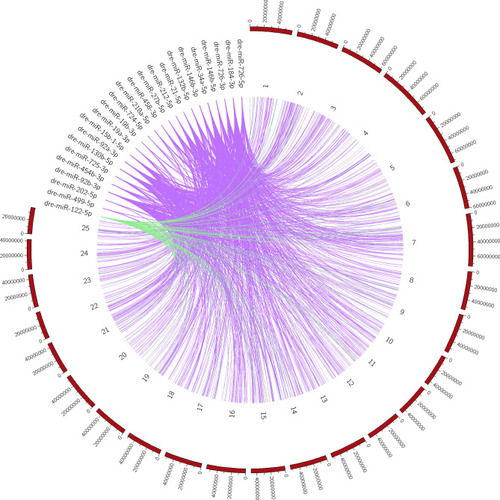
Circular localization of predicted target genes from differentially expressed (DE) miRNAs in the zebrafish genome. 407 predicted target genes of DE miRNAs in the ovary (purple) and 85 predicted target genes in the testis (green) were distributed throughout the zebrafish genome, with the highest percentage present in chromosomes 7 and 14 for ovary and testis, respectively. |

Chromosomal distribution in the zebrafish genome of the number of predicted target genes obtained from differentially expressed miRNAs after high temperature in the ovary ( |

Fluorescent in situ hybridization (FISH) of dre-miR-146b-5p in the ovary of adult zebrafish. A total of 6 female fish were used to obtain the results. ( |
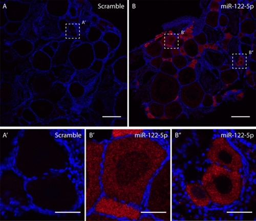
Fluorescent in situ hybridization (FISH) of dre-miR-122-5p in the ovary of adult zebrafish. A total of 6 female fish were used to obtain the results. Sections |