
- Title
-
Hyperactivation of Wnt/β-catenin and Jak/Stat3 pathways in human and zebrafish foetal growth restriction models: Implications for pharmacological rescue
- Authors
- Risato, G., Celeghin, R., Brañas Casas, R., Dinarello, A., Zuppardo, A., Vettori, A., Pilichou, K., Thiene, G., Basso, C., Argenton, F., Visentin, S., Cosmi, E., Tiso, N., Beffagna, G.
- Source
- Full text @ Front Cell Dev Biol
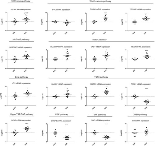
Signalling pathway dysregulation in human FGR samples. Quantitative RT-PCR analysis performed on umbilical cords from FGR cases and AGA controls, focusing on ten signalling pathways, identified significant upregulation in seven pathways (Hif/Hypoxia, Wnt/β-catenin, Jak/Stat3, Notch, Bmp, TGFβ, Hippo/YAP-TAZ), downregulation in two (FGF, Shh) and no modification in one (CREB). Sample size: |

Long-term hypoxia treatment leads to growth restriction in zebrafish embryos. |

Long-term hypoxia induces cardiovascular modifications in zebrafish embryos/larvae. |
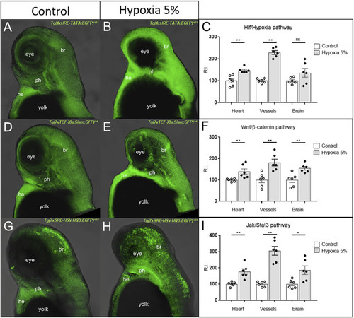
Hyper-activation of Hif/Hypoxia, Wnt/β-catenin and Jak/Stat3 signalling in the cardiovascular and brain regions of hypoxia-treated zebrafish. The GFP-based (green) reporters for Hif/Hypoxia |

Stat3 signalling modulation does not rescue FGR anomalies. Downregulation of Stat3 signalling with 50 μM AG490 inhibitor does not rescue body length, eye size and cardiac morphology under hypoxia. Upregulation of Stat3 signalling with 20 μM LIF agonist does not rescue body length, eye size and cardiac morphology under hypoxia. All embryos, at 3 dpf, are displayed in lateral view, anterior to the left. Sample size: |
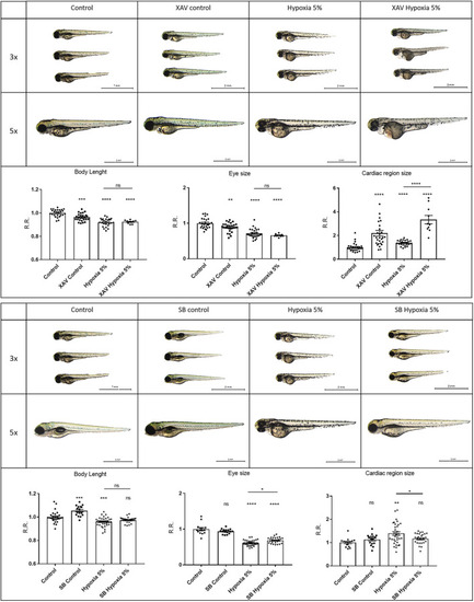
Wnt/β-catenin signalling upregulation can rescue FGR anomalies. Downregulation of Wnt/β-catenin signalling with 5 µM XAV939 (XAV) inhibitor does not rescue body length or eye size, and worsens the pericardial effusion under hypoxia. Upregulation of Wnt/β-catenin signalling with 40 µM SB216763 (SB) agonist can rescue the body length and the pericardial effusion under hypoxia. All embryos, at 3 dpf, are displayed in lateral view, anterior to the left. For display purposes, images of control embryos under normoxia and hypoxia correspond to control images of |

Wnt/β-catenin upregulation can rescue hypoxia-induced vascular defects. Upregulation of Wnt/β-catenin signalling using 40 µM SB216763 (SB) can rescue the vascular organization (reciprocal distance and vessel diameter) in hypoxia-treated embryos. Conversely, downregulation of Wnt/β-catenin signalling using 5 µM XAV939 leads to a worsened phenotype. Intersegmental vessels (arrows) were inspected in 3 dpf embryos, after 2-days long incubation in normoxia (control) or 5% hypoxia, exploiting the endothelial-specific transgene |