
- Title
-
Aquaporin 8ab is required in zebrafish embryonic intestine development
- Authors
- Wang, S., Qin, Y., Sheng, J., Duan, X., Shen, L., Liu, D.
- Source
- Full text @ Acta. Biochim. Biophys. Sin (Shanghai)
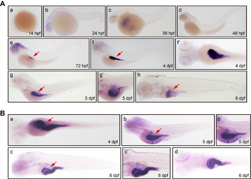
(A) Whole mount EXPRESSION / LABELING:
|
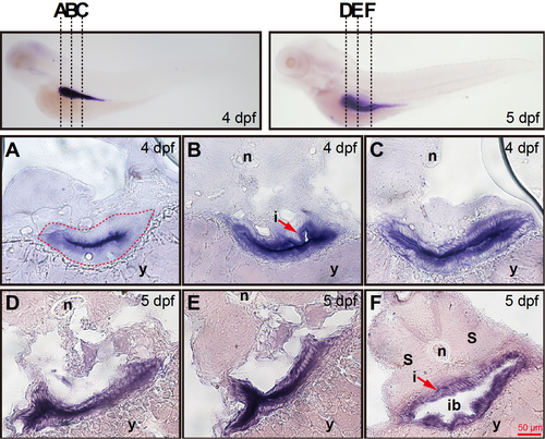
(A–C) Corresponding transverse sections through the three different regions of intestine at 4 dpf depicted in left image of the top row. (A) The |

(A) Schematic diagram showing |
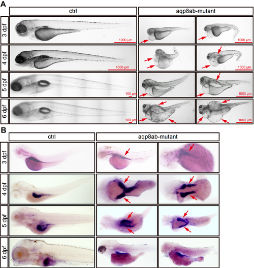
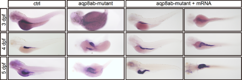
(A) Microscopy analysis of embryos development at 3–6 dpf in control group and EXPRESSION / LABELING:
PHENOTYPE:
|
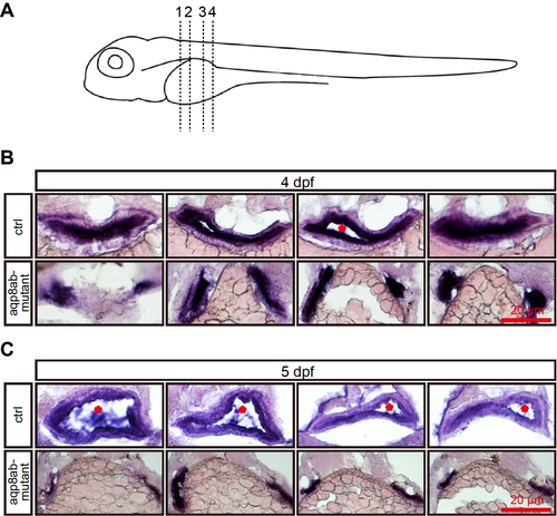
(A) Schematic diagram showing transverse sections through the trunk at four different regions. (B) At 4 dpf, the control group showed a single intestinal tract, in which a lumen was already formed (stars). The |
At 4–5 dpf, the control group had a single intestinal tract, the EXPRESSION / LABELING:
PHENOTYPE:
|