
- Title
-
Dysregulated myosin in Hermansky-Pudlak syndrome lung fibroblasts is associated with increased cell motility
- Authors
- Imani, J., Bodine, S.P.M., Lamattina, A.M., Ma, D.D., Shrestha, S., Maynard, D.M., Bishop, K., Nwokeji, A., Malicdan, M.C.V., Testa, L.C., Sood, R., Stump, B., Rosas, I.O., Perrella, M.A., Handin, R., Young, L.R., Gochuico, B.R., El-Chemaly, S.
- Source
- Full text @ Respir Res
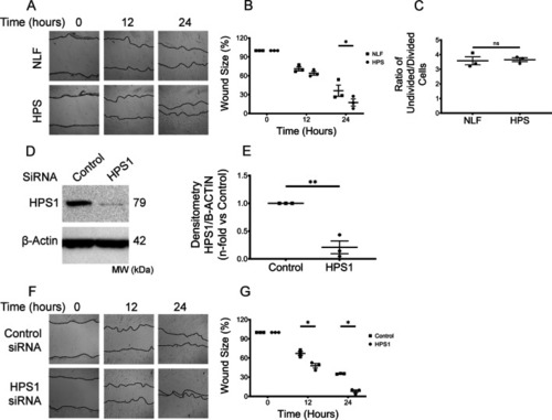
Enhanced HPSLF migration in vitro is HPS1-dependent. A, F Representative microscopy images of scratch assays at 0, 12, and 24 h after gaps were generated. Scratch assays were performed with A NLF (n = 3 technical replicates) and HPSLF (n = 3 technical replicates); F NLF transfected with control or HPS1 siRNA; B, G Quantitative analysis of the migration potential of B NLF and HPSLF; G NLF transfected with control or HPS1 siRNA. The rate of fibroblast migration was determined by calculating the percentage of open wound area at the indicated time points to that of the corresponding initial scratch. C Ratio of CFSE MFI from proliferating cells over undivided cells D Western blot analysis of HPS1 protein expression in NLF transfected with either control or HPS1 siRNA. β-Actin was used as a loading control. E Ratio of HPS1 to β-Actin density expressed as fold change relative to NLF transfected with control siRNA. Data are expressed as mean ± SEM of three independent experiments. B, G Data analyzed using two-way mixed ANOVA *P < 0.05. C, E Data was analyzed using Student’s t-test *P < 0.05, **P < 0.01 |
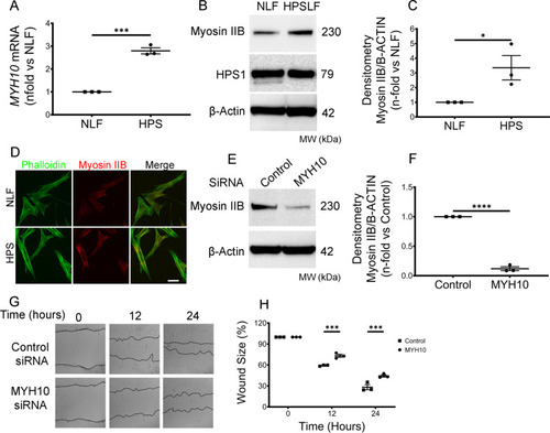
Enhanced migration of HPSLF is Myosin IIB dependent. A Real-time PCR analysis of MYH10 mRNA in NLF (n = 3 technical replicates) and HPSLF (n = 3 technical replicates). Results were expressed as fold change relative to NLF. B Western blot analysis of Myosin IIB and HPS1. Protein expression in control NLF and HPSLF cells are shown; β-Actin was used as a loading control. C Ratio of Myosin IIB to β-Actin density expressed as fold-change relative to control NLF. D Immunofluorescent images of NLF and HPSLF stained with Phalloidin (green) and Myosin IIB (red). E Western blot analysis of Myosin IIB protein expressions in HPSLF transfected with either control or MYH10 siRNA. β-Actin was used as a loading control. F Ratio of Myosin IIB to β-Actin density expressed as fold-change relative to control siRNA. G Representative microscopy images of scratch assays at 0, 12, and 24 h after gaps were generated in control or MYH10 siRNA transfected HPSLF. H Quantitative analysis of the migration potential of control or MYH10 siRNA transfected HPSLF. The rate of fibroblast migration was determined by calculating the percentage of open wound area at the indicated time points to that of the corresponding initial scratch. Data are expressed as mean ± SEM of three independent experiments. A, B, E Data were analyzed using students t-test *P < 0.5, ***P < 0.001, ****P < 0.0001. G Data were analyzed using two-way mixed ANOVA ***P < 0.001 |
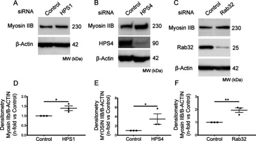
A BLOC3-dependent mechanism modulates Myosin IIB level in HPSLF. A and D Western blot analysis of Myosin IIB protein expression in NLF (n = 3 technical replicates) transfected with either control or HPS1 siRNA. β-Actin was used as a loading control (same membranes as in Fig. 1D). Ratio of Myosin IIB to β-Actin density expressed as fold change relative to NLF transfected with control siRNA. B and E Western blot analysis of Myosin IIB protein expression in NLF (n = 3 technical replicates) transfected with either control or HPS4 siRNA. β-Actin was used as a loading control. Ratio of Myosin IIB to β-Actin density expressed as fold change relative to NLF transfected with control siRNA. C and F Western blot analysis of Myosin IIB in NLF (n = 3 technical replicates) transfected with either control or Rab32 siRNA. β-Actin was used as a loading control. The ratio of Myosin IIB to β-Actin density expressed as fold change relative to NLF transfected with control siRNA. Data are expressed as mean ± SEM of three independent experiments. Data were analyzed using a student’s t-test *P < 0.05, **P < 0.01, ****P < 0.0001 |
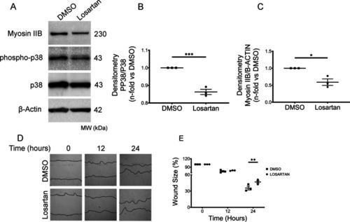
Losartan blocks the enhanced HPSLF migration in vitro. A Western blot analysis of Myosin IIB, phospho-p38, and p38 protein expression in HPSLF (n = 3 technical replicates) treated with vehicle (DMSO) or with losartan (100 nM). β-Actin was used as a loading control. B Ratio of phospho-p38 to p38 density and C Myosin IIB to β-Actin density expressed as fold-change relative to DMSO. D Representative microscopy images of scratch assays at 0, 12, and 24 h after gaps were generated. Scratch assays were performed with HPSLF (n = 3 technical replicates) treated with vehicle (DMSO) or losartan (100 nM). E Quantitative analysis of the migration potential. The rate of fibroblast migration was determined by calculating the percentage of open wound area at the indicated time points to that of the corresponding initial scratch. Data are expressed as mean ± SEM of three independent experiments. B, C Data analyzed using students t-test *P < 0.5, ***P < 0.001. E Data analyzed using two-way mixed ANOVA **P < 0.01 |
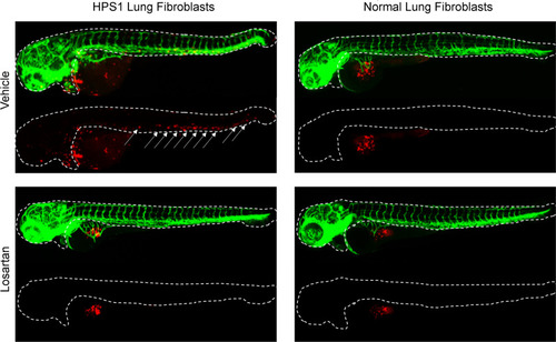
Modulation of enhanced migratory capacity of HPS lung fibroblasts (HPSLF) by losartan in vivo. HPSLF and normal lung fibroblasts (NLF) were treated with losartan or vehicle (PBS) and labeled with a CM-Dil live-cell marker (red). 75 cells were injected into the yolk sac of 48 h post-fertilization Tg(fli1:GFP, green) zebrafish embryos expressing GFP in their blood vessels. HPSLFs exhibit stronger migratory capacity than NLFs at 24 h (not shown) and 48 h after injection. Arrows indicate the migration of labeled fibroblasts from the yolk sac into outlined regions of zebrafish embryos. Treatment with losartan reduces HPSLF migratory capacity compared to baseline |