FIGURE SUMMARY
Title

Profilin 2 and Endothelial Exosomal Profilin 2 Promote Angiogenesis and Myocardial Infarction Repair in Mice

Authors
Li, Z., Huo, X., Chen, K., Yang, F., Tan, W., Zhang, Q., Yu, H., Li, C., Zhou, D., Chen, H., Zhao, B., Wang, Y., Chen, Z., Du, X.
Source
Full text @ Front Cardiovasc Med

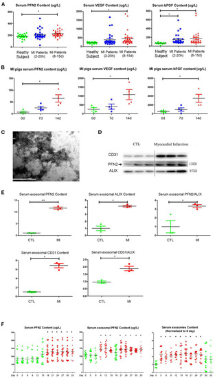
Increased PFN2 and exosomal PFN2 levels in the serum of patients with MI and in animal models. (A) Enhanced PFN2 levels, VEGFA and bFGF levels in patients with MI (patients with MI 2–20 h, n = 30; MI 8–15 d, n = 30; CTL, n = 24). (B) Enhanced PFN2 levels, VEGFA and bFGF levels in pigs with MI (pigs with MI, n = 4; CTL, n = 4). (C) Identification of serum exosomes in patients with MI by electron microscopy. (D) Exosomal PFN2 between CTL (n = 3) and MI groups (n = 3). (E) Exosomal PFN2 levels between CTL (n = 3) and MI groups (n = 3) (The protein level was normalized to control). (F) Serum PFN2 levels, Exosomal PFN2 levels and Exosomes in rats (n = 13) before and after MI. *p < 0.05, one-way ANOVA and repeated-measures ANOVA (Tukey).

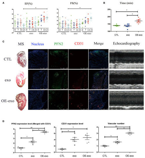
Exosomal PFN2 repairs infarction in mouse with MI and restores cardiac function. (A) The cardiac function of mouse with MI as evaluated by type-B ultrasonic test EF and FS after treatment with exosomes demonstrated the benefit to MI impairment. (B) Physical fitness was evaluated by fatigue test after treatment with exosomes in mouse with MI which displayed significant improvement of heart function, n = 7. (C) Infarction volume (Masson's staining) decreased and PFN2 (green)/CD31 (red) staining increased in heart tissue of mouse with MI after treatment with exosomes. (D) Statistical results of (C). n = 3, normalized to control. *p < 0.05, one-way ANOVA (Tukey).

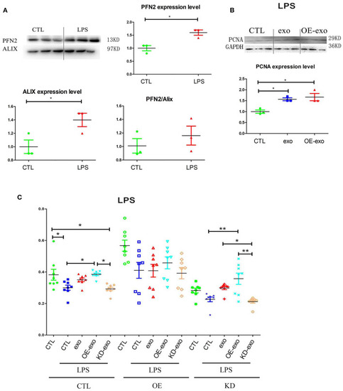
PFN2 protects ECs after LPS treatment. (A) PFN2 and Alix expression levels in LPS-treated EC exosomes, n = 3, normalized to control. (B) PCNA expression levels of ECs under LPS stimulus after treatment with various exosomes, n = 3, normalized to control. (C) Viability of ECs under LPS stimulation after treatment with various exosomes. n = 8. *p < 0.05, one-way ANOVA (Tukey).

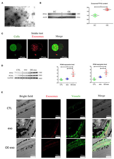
Exosomal PFN2 enhances the proliferation ability of ECs and OE-exo treated zebrafish showed significantly increased vessel number than exo and control. (A) The exosomes secreted from ECs were identified by electron microscopy. (B) PFN2 expression in exosomes (n = 3, the expression levels were normalized to control). (C) Exosomes could be internalized by ECs. (D) PFN2 and PCNA expression levels in CTL, exosomes from normal ECs (exo), PFN2-overexpressing ECs (OE-exo), and PFN2 knockdown ECs (KD-exo), n = 3, and the expression levels were normalized to control. (E) OE-exo-treated zebrafish showed significantly increased vessel number than exo and control. Exo, exosomes from normal ECs; OE-exo, exosomes from PFN2-overexpressing ECs; CTL, PBS as control. *p < 0.05, one-way ANOVA (Tukey).

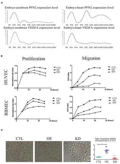
PFN2 promotes proliferation and migration of ECs, as well as tube formation and reaches the maximum level in 10-day embryo. (A) The mRNA levels of PFN2 and VEGFA at different developmental stages of gerbils (n = 15) embryo (prenatal), brain and heart (postnatal). (B) PFN2 significantly promoted proliferation of HUVECs (n = 3) and RBMECs (n = 3), as well as showed a positive effect on migration of HUVECs (n = 3) and RBMECs (n = 3). (C) The tube length in PFN2-overexpressing ECs (OE) was significantly increased than that in blank CTL and PFN2 knockdown ECs (KD), n = 3. *p < 0.05, one-way ANOVA and repeated-measures ANOVA (Tukey).

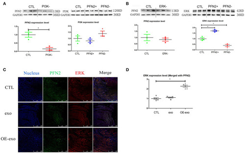
PFN2 involved molecular signaling pathway. (A) PFN2 expression level in HUVECs treated with PI3K inhibitor and PI3K expression level in OE and KD, n = 3, normalized to control. (B) PFN2 expression level in HUVECs treated with ERK inhibitor and ERK expression level in OE and KD, n = 3, normalized to control. (C) ERK and PFN2 expression levels in the heart tissue of MI mice treated with different exosomes, n = 3, normalized to control. (D) Statistical result of (C). *p < 0.05, Student's t-test and one-way ANOVA (Tukey).

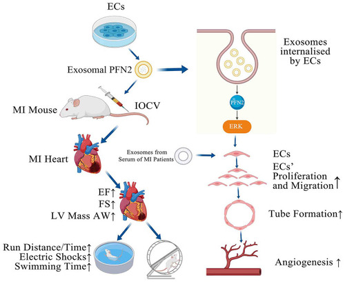
GRAPHICAL ABSTRACT. PFN2-overexpressing ECs (OE-exo) treated MI mice showed improvement in infarction volume, cardiac function and motor ability, and PFN2/ OE-exo significantly enhanced EC proliferation, migration, tube formation ability and angiogenesis.

Acknowledgments
This image is the copyrighted work of the attributed author or publisher, and ZFIN has permission only to display this image to its users. Additional permissions should be obtained from the applicable author or publisher of the image. Full text @ Front Cardiovasc Med