
- Title
-
A new transgenic reporter line reveals expression of protocadherin 9 at a cellular level within the zebrafish central nervous system
- Authors
- Habicher, J., Manuel, R., Pedroni, A., Ferebee, C., Ampatzis, K., Boije, H.
- Source
- Full text @ Gene Expr. Patterns
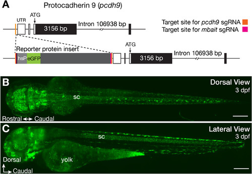
Generation of a pcdh9:hs:eGFP reporter line. Targeted genomic integration of the eGFP reporter construct (A) and overview of eGFP expression in Tg(pcdh9:hs:eGFP) larvae at 3 days post fertilisation (dpf) in a dorsal (B) and lateral (C) view of the brain and spinal cord (sc). Scale bars equal 200 μm. EXPRESSION / LABELING:
Construct:
Tg5(hsp70l:EGFP)
|
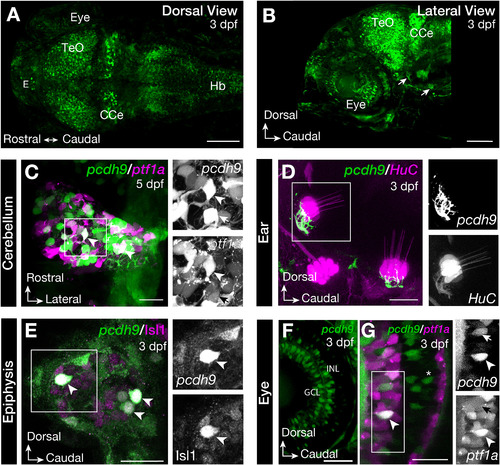
Tg(pcdh9:hs:GFP) expression in the brain of zebrafish larvae. Dorsal (A) and lateral view (B) of the brain of Tg(pcdh9:hs:GFP) zebrafish at 3 days post fertilisation (dpf) show expression in the optic tectum (TeO), cerebellum (CCe), epiphysis (E), in the retina of the eye, the hindbrain (Hb) and axons connecting to the hair cells of the ear (arrow). In the cerebellum pcdh9 was expressed in a subpopulation of Purkinje cells labelled by Tg(ptf1a:dsRed) (arrowhead), ptf1a negative cells are indicated with an arrow (C). In the ear, pcdh9 positive axons connecting to the hair cells, labelled by Tg(HuC:GAL4; UAS:RFP), were observed (D). Immunohistochemistry showed that all pcdh9-eGFP positive cells in the epiphysis were positive for Isl1 (E). The pcdh9-eGFP positive cells in the retina were located in the ganglion cell layer (GCL) and in the inner nuclear layer (INL) (F). In the INL a subset of pcdh9 positive cells were amacrine cells labelled by Tg(ptf1a:dsred) (arrowhead), a subset negative for ptf1a (arrows). Bipolar cells were also pcdh9 positive (G, asterix). Scale bars equal 100 μm in A-B and 20 μm in C-G. EXPRESSION / LABELING:
|
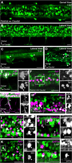
Tg(pcdh9:hs:GFP) zebrafish larvae in the zebrafish spinal cord. Dorsal (A) and lateral view (B) of the spinal cord of Tg(pcdh9:hs:GFP) zebrafish at 3 days post fertilisation (dpf) showed pcdh9 positive cells within and outside the spinal cord. One cell of the dorsal root ganglia per hemi-segment was pcdh9 positive at 6 dpf (C; arrowhead) and up to 10 cells per hemi-segment were labelled at 19 dpf (D; arrowheads). These cells were Isl1 positive (E). Isl1 also stains the Rohon Beard cells, which were negative for pcdh9 (F). Crossing Tg(pcdh9:hs:eGFP) with Tg(mnx1:Gal4; UAS:RFP) revealed the pcdh9 positive glia cells encasing the motor neuron axons leaving the spinal cord (G) and overlap of mnx1 and pcdh9 expressing cells in secondary motor neurons (H; arrowhead). Immunohistochemistry for Evx2 revealed overlap with pcdh9-eGFP positive interneurons in the spinal cord (I). Expression of pcdh9 was also observed in the dI6 interneurons marked by dmrt3a (Tg(dmrt3a:GAL4; UAS:RFP)) (J) or Wt1 (immunohistochemistry) (K), but not in the dI4 interneurons labelled by (Tg(ptf1a:dsred) (L). Scale bars equal 20 μm. EXPRESSION / LABELING:
|

Tg(pcdh9:hs:GFP) juvenile zebrafish exhibit expression both in the spinal cord and in the cerebellum. All the pcdh9 positive cells expressed the pan-neuronal marker HuC/D (A). Retrograde labelling of motor neurons showed a specific expression of pcdh9 in a portion of slow secondary motor neurons but not in primary, or fast and intermediate secondary motor neurons (B, C). In the cerebellum, pcdh9 positive cells were positive for ZebrinII both in the valvula lateralis (Val) and valvula medialis (Vam) (D) and in the corpus cerebelli (CCe) (E). Scale bars equal 20 μm. EXPRESSION / LABELING:
|

ZFIN is incorporating published figure images and captions as part of an ongoing project. Figures from some publications have not yet been curated, or are not available for display because of copyright restrictions. EXPRESSION / LABELING:
|
|
ZFIN is incorporating published figure images and captions as part of an ongoing project. Figures from some publications have not yet been curated, or are not available for display because of copyright restrictions. EXPRESSION / LABELING:
|
Reprinted from Gene expression patterns : GEP, 44, Habicher, J., Manuel, R., Pedroni, A., Ferebee, C., Ampatzis, K., Boije, H., A new transgenic reporter line reveals expression of protocadherin 9 at a cellular level within the zebrafish central nervous system, 119246, Copyright (2022) with permission from Elsevier. Full text @ Gene Expr. Patterns