
Fig. 3
- ID
- ZDB-IMAGE-220608-5
- Genes
- Antibodies
- Publication
- Habicher et al., 2022 - A new transgenic reporter line reveals expression of protocadherin 9 at a cellular level within the zebrafish central nervous system
- All Figures
- Figures for Habicher et al., 2022
|
Fig. 3
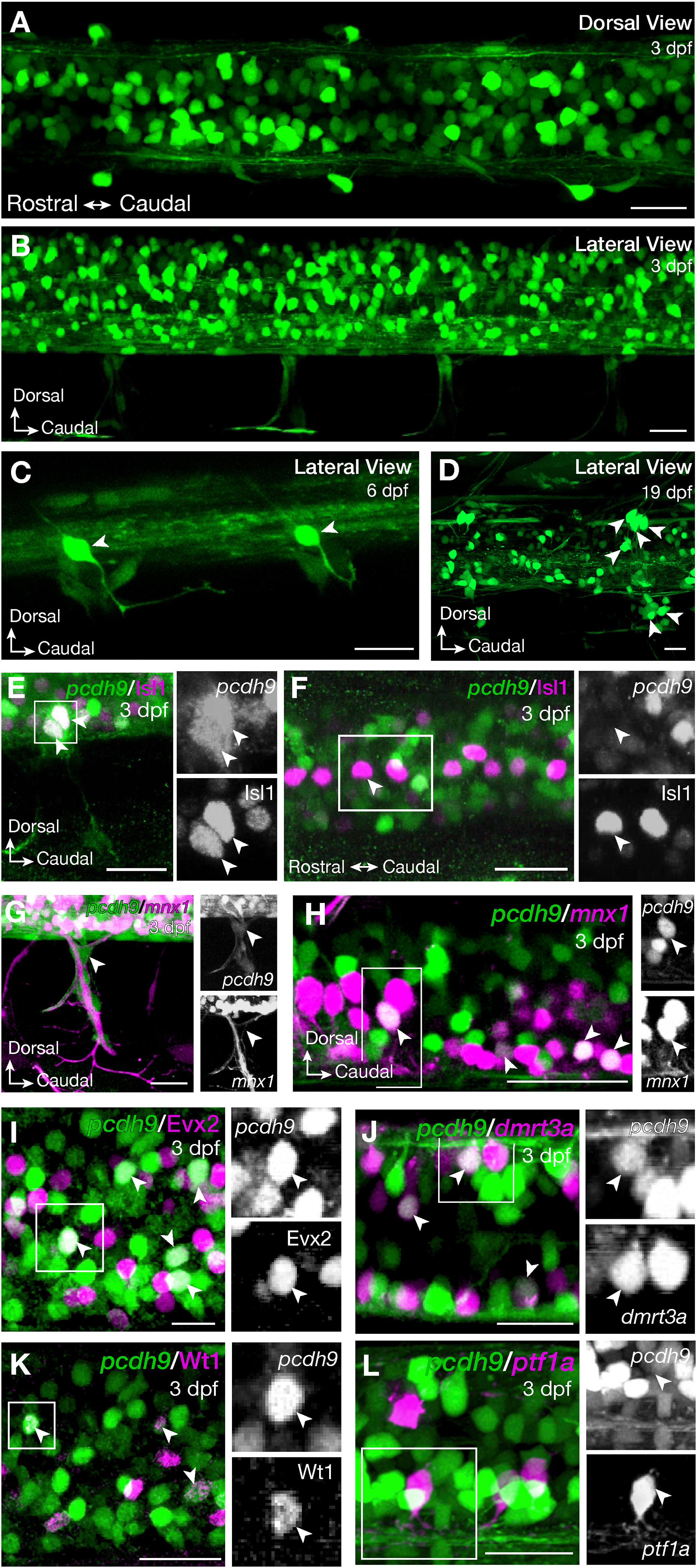
Tg(pcdh9:hs:GFP) zebrafish larvae in the zebrafish spinal cord. Dorsal (A) and lateral view (B) of the spinal cord of Tg(pcdh9:hs:GFP) zebrafish at 3 days post fertilisation (dpf) showed pcdh9 positive cells within and outside the spinal cord. One cell of the dorsal root ganglia per hemi-segment was pcdh9 positive at 6 dpf (C; arrowhead) and up to 10 cells per hemi-segment were labelled at 19 dpf (D; arrowheads). These cells were Isl1 positive (E). Isl1 also stains the Rohon Beard cells, which were negative for pcdh9 (F). Crossing Tg(pcdh9:hs:eGFP) with Tg(mnx1:Gal4; UAS:RFP) revealed the pcdh9 positive glia cells encasing the motor neuron axons leaving the spinal cord (G) and overlap of mnx1 and pcdh9 expressing cells in secondary motor neurons (H; arrowhead). Immunohistochemistry for Evx2 revealed overlap with pcdh9-eGFP positive interneurons in the spinal cord (I). Expression of pcdh9 was also observed in the dI6 interneurons marked by dmrt3a (Tg(dmrt3a:GAL4; UAS:RFP)) (J) or Wt1 (immunohistochemistry) (K), but not in the dI4 interneurons labelled by (Tg(ptf1a:dsred) (L). Scale bars equal 20 μm.
Reprinted from Gene expression patterns : GEP, 44, Habicher, J., Manuel, R., Pedroni, A., Ferebee, C., Ampatzis, K., Boije, H., A new transgenic reporter line reveals expression of protocadherin 9 at a cellular level within the zebrafish central nervous system, 119246, Copyright (2022) with permission from Elsevier. Full text @ Gene Expr. Patterns