
- Title
-
Compression Fractures and Partial Phenotype Rescue With a Low Phosphorus Diet in the Chihuahua Zebrafish Osteogenesis Imperfecta Model
- Authors
- Cotti, S., Huysseune, A., Larionova, D., Koppe, W., Forlino, A., Witten, P.E.
- Source
- Full text @ Front Endocrinol (Lausanne)
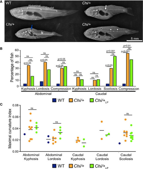
Vertebral column deformities in PHENOTYPE:
|
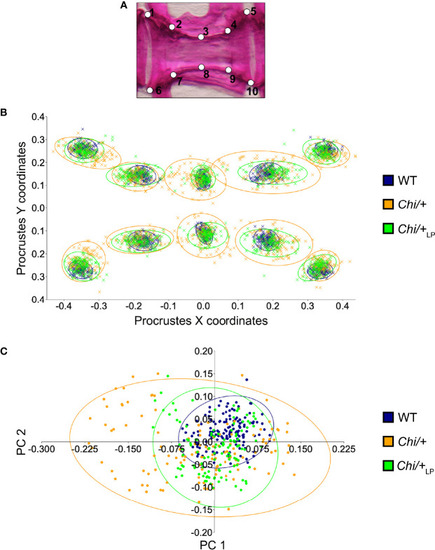
Vertebral body shape variation in PHENOTYPE:
|
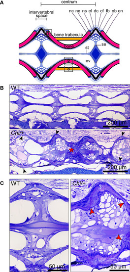
Histology of vertebral column of WT, PHENOTYPE:
|
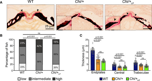
Figure 4 Chi/+ vertebral bone structures are thin and highly mineralised. The LP diet restores the osteoid. (A) Sagittal histological non-demineralised sections stained with Von Kossa/Van Gieson show that three months old Chi/+ animals, compared to WT animals, have highly mineralised endplates. No osteoid layer can be identified. The osteoid (pink, black arrowheads) is restored in Chi/+LP. Mineralised bone: black; dense collagen and non-mineralised bone: red. (B) Quantitative analysis of vertebral body endplate mineralisation (scored as low, intermediate or high) based on whole mount-stained specimens shows that Chi/+ animals exhibit a higher degree of mineralisation compared to WT animals. The LP diet reduces mineralisation of the vertebral body endplates in some Chi/+ individuals. The first 5 caudal vertebral centra in WT (n=15), Chi/+ (n=13) and Chi/+LP (n=12) were analysed. Chi-square test followed by Bonferroni correction, p values are indicated, ns: non-significant. (C) Measurements of bone structure thickness at three locations: (i) vertebral endplates, (ii) central region of vertebrae and (iii) trabecular bone (see Figure 3A for locations). Compared to WT animals, Chi/+ and Chi/+LP animals have thinner bone structures in all three locations (see also Table 2). Thickness of bone structures was measured on toluidine blue stained sections in 5 to 10 vertebral centra in WT (n=4), Chi/+ (n=4) and Chi/+LP (n=5). Mann-Whitney test followed by Bonferroni correction, p values are indicated, ns: non-significant. PHENOTYPE:
|
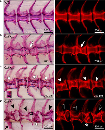
Different grades of PHENOTYPE:
|
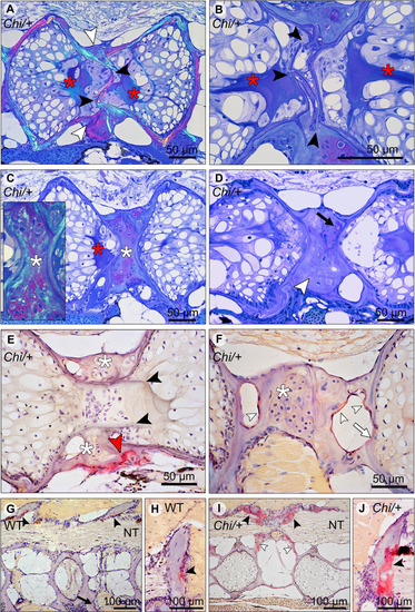
Compression fractures and bone resorption in PHENOTYPE:
|
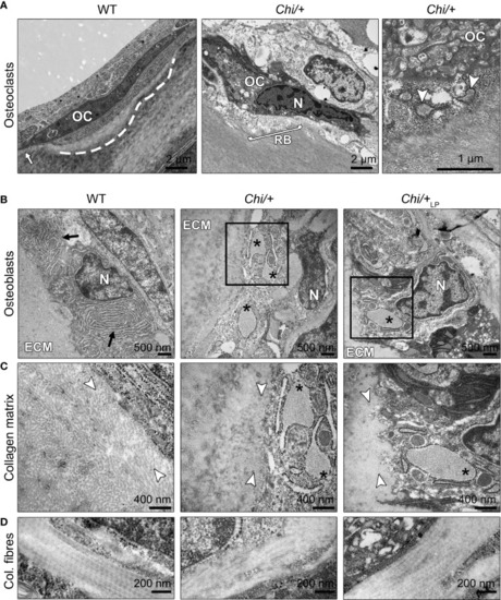
Ultrastructure of bone cells and bone matrix in |