
- Title
-
Involvement of the zebrafish trrap gene in craniofacial development
- Authors
- Suzuki, T., Hirai, Y., Uehara, T., Ohga, R., Kosaki, K., Kawahara, A.
- Source
- Full text @ Sci. Rep.

ZFIN is incorporating published figure images and captions as part of an ongoing project. Figures from some publications have not yet been curated, or are not available for display because of copyright restrictions. PHENOTYPE:
|
|
ZFIN is incorporating published figure images and captions as part of an ongoing project. Figures from some publications have not yet been curated, or are not available for display because of copyright restrictions. EXPRESSION / LABELING:
|
|
ZFIN is incorporating published figure images and captions as part of an ongoing project. Figures from some publications have not yet been curated, or are not available for display because of copyright restrictions. EXPRESSION / LABELING:
|
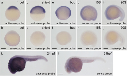
Expression of the |
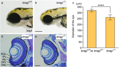
Small eyes in the |

Small heads in the PHENOTYPE:
|
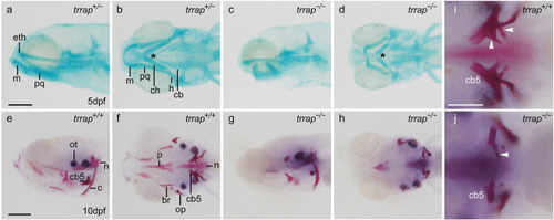
Morphological defects in the pharyngeal arches and teeth of the |
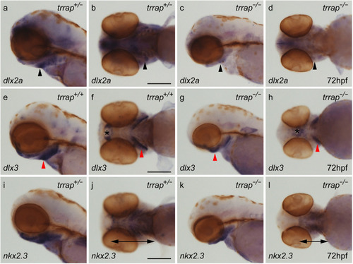
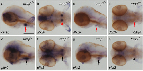
Differential expression of pharyngeal arch genes in the EXPRESSION / LABELING:
PHENOTYPE:
|
Differential expression of tooth marker genes in the EXPRESSION / LABELING:
PHENOTYPE:
|