
- Title
-
A lysosome-targeted dextran-doxorubicin nanodrug overcomes doxorubicin-induced chemoresistance of myeloid leukemia
- Authors
- Zeng, Y., Zhang, X., Lin, D., Feng, X., Liu, Y., Fang, Z., Zhang, W., Chen, Y., Zhao, M., Wu, J., Jiang, L.
- Source
- Full text @ J. Hematol. Oncol.
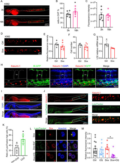
Hypoxic CHT harbored chemoresistant leukemic cells with increased lysosomes that sequestered Dox to prevent its nuclear entry and cytotoxicity. A–C K562 cells were microinjected into 2dpf embryos. The cell number and fluorescent intensity of leukemic cells localized in CHT at different time points post-injection were counted. D–G K562-xenografted zebrafish were treated with Dox from one-day-post injection (1 dpi) to 3 dpi, and the leukemic cells in CHT were quantified with the fluorescent intensity (E), the cell number (F) and the mRNA expression of human ribosome gene L32 (G). H Dox-resistant Kasumi-1 cells were mainly localized in the CVP vessels visualized by flk:GFP fish. CVP-caudal vein plexus, CA-caudal aorta, ISV-intersegmental vessel. I The 2dpf zebrafish embryos were stained with the PIM antibody to detect hypoxia tissue. The CHT was labeled with yellow rectangles. J, K The Kasumi-1-xenografted zebrafish were stained with LysoTracker and quantified for lysosome enrichment in CHT and non-CHT in vivo. The entire CHT in the left panel was labeled with yellow rectangles, and the region in blue rectangles were magnified in the right panels to show details. L The subcellular localization of Dox in K562 cells was visualized by its autonomous red fluorescence in Dox, Dox + Baf or Dox + CQ. M The chemoresistant K562 cells that localized in CHT were more sensitive to Dox + CQ while CQ alone has no toxicity. Bar plots are shown as average ± SEM. The statistical significance between groups was determined using Student’s t-test or ANOVA analysis. *Indicates p < 0.05; **p < 0.01; ***p < 0.001; ****p < 0.0001 |
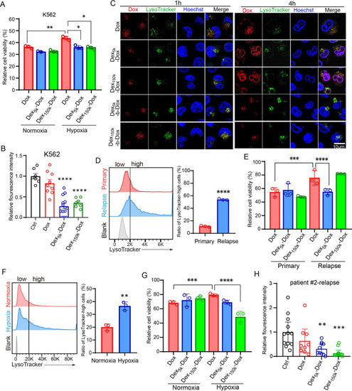
The pH-sensitive Dex-Dox nanomedicine promoted the nuclear entry and cytotoxicity of Dox in hypoxic leukemic cells to overcome chemoresistance. A The hypoxia-cultured K562 cells have higher viability post Dox treatment, but the viability was significantly reduced in Dex5k/150k-Dox. B The K562-xenografted-zebrafish embryos were treated with Dox or Dex5k/150k-Dox, and leukemic cells in CHT were counted at two days post-treatment. The results showed that Dex5k/150k-Dox had much higher toxicity against chemoresistant leukemic cells. C The subcellular localization of Dox was analyzed by its autonomous red fluorescence in Dox, Dex-Dox or Dex-b-Dox treated K562 cells. D, E The bone marrow specimen from the same patient (#1) at primary or relapsed stage were collected for staining with LysoTracker (D) or cell counting after treatment with Dox or Dex5k/150k-Dox (E). F, G The bone marrow specimen from the relapsed patient #2 was collected and incubated in hypoxia to measure the ratio of LysoTracker-high cells (F) and count the cell number after treatment with Dox or Dex5k/150k-Dox (G). H The zebrafish embryos were xenografted with the relapsed leukemic cells from patient #2 and treated with Dox or Dex5k/150k-Dox. The fluorescent intensity of leukemic cells in CHT was quantified at two days post-treatment. Bar plots are shown as average ± SEM. The statistical significance between groups was determined using Student’s t-test or ANOVA analysis. * Indicates p < 0.05; **p < 0.01; ***p < 0.001; ****p < 0.0001 |