
|
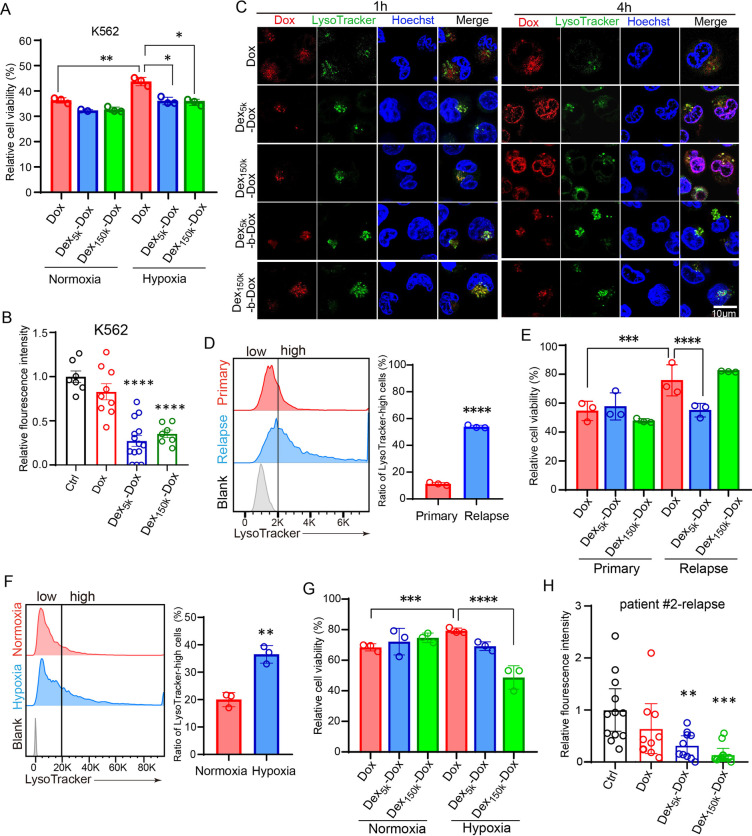
Fig. 2
The pH-sensitive Dex-Dox nanomedicine promoted the nuclear entry and cytotoxicity of Dox in hypoxic leukemic cells to overcome chemoresistance. A The hypoxia-cultured K562 cells have higher viability post Dox treatment, but the viability was significantly reduced in Dex5k/150k-Dox. B The K562-xenografted-zebrafish embryos were treated with Dox or Dex5k/150k-Dox, and leukemic cells in CHT were counted at two days post-treatment. The results showed that Dex5k/150k-Dox had much higher toxicity against chemoresistant leukemic cells. C The subcellular localization of Dox was analyzed by its autonomous red fluorescence in Dox, Dex-Dox or Dex-b-Dox treated K562 cells. D, E The bone marrow specimen from the same patient (#1) at primary or relapsed stage were collected for staining with LysoTracker (D) or cell counting after treatment with Dox or Dex5k/150k-Dox (E). F, G The bone marrow specimen from the relapsed patient #2 was collected and incubated in hypoxia to measure the ratio of LysoTracker-high cells (F) and count the cell number after treatment with Dox or Dex5k/150k-Dox (G). H The zebrafish embryos were xenografted with the relapsed leukemic cells from patient #2 and treated with Dox or Dex5k/150k-Dox. The fluorescent intensity of leukemic cells in CHT was quantified at two days post-treatment. Bar plots are shown as average ± SEM. The statistical significance between groups was determined using Student’s t-test or ANOVA analysis. * Indicates p < 0.05; **p < 0.01; ***p < 0.001; ****p < 0.0001