FIGURE SUMMARY
Title

The RNA-binding protein Igf2bp3 is critical for embryonic and germline development in zebrafish

Authors
Vong, Y.H., Sivashanmugam, L., Leech, R., Zaucker, A., Jones, A., Sampath, K.
Source
Full text @ PLoS Genet.

Loss of <italic toggle='yes'>igf2bp3</italic> results in skewed male:female sex ratios.

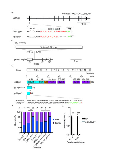
A. Generation of the igf2bp3 mutant allele by Cas9. Schematic showing the igf2bp3 locus on chromosome 19. Exon 1 of igf2bp3 was targeted by CRISPR-Cas9, resulting in a 7 bp deletion and frameshift. B. Schematic of the igf2bp3la659Tg with a 6 kb Tg(nLacZ-GTvirus) retroviral insertion in intron 1. C. The igf2bp3Δ7 mutant is predicted to be protein null. The igf2bp3Δ7 mutation generates a premature stop codon at residue 46 whereas the igf2bp3la659Tg results in a predicted truncated protein of 65 amino acids. D. Loss of igf2bp3 results in skewed sex ratios. Adult zygotic igf2bp3Δ7 and MZigf2bp3la659Tg mutant fish show a significant male bias compared to wild type, heterozygous igf2bp3Δ7 as well as igf2bp3la659Tg, and Zigf2bp3la659Tg fish. Asterisks indicate level of significance (p *<0.05, **<0.01, ***<0.001). E. The igf2bp3Δ7 mutation results in reduced igf2bp3 levels. qRT-PCRs at the 1-cell and 4-cell stages show significantly reduced igf2bp3 transcript levels in igf2bp3Δ7 mutant embryos (p<0.001).

Maternal <italic toggle='yes'>igf2bp3</italic> is required for normal embryonic development.

A. Maternal igf2bp3Δ7 mutants show severe defects by blastula and gastrula stages. B. At the 64-cell stage, most maternal igf2bp3Δ7 mutants are similar to wild type embryos (left), but a proportion of embryos (right; <15%) show rounded cells (red arrowheads). C-D. The majority of igf2bp3 mutants do not survive beyond 1 dpf. E. Animal-vegetal markers (gdf3, dazl and wnt8a) show altered distribution in igf2bp3Δ7 embryos by whole mount in situ hybridization (WISH). F. qRT-PCRs show that expression levels of gdf3, dazl and wnt8a transcripts is not significantly different from wild type control embryos at the 1-cell stage. G-I. qRT-PCR with maternal igf2bp3Δ7 embryos show that expression of igf2bp3 transcripts is significantly reduced at all stages examined (1 cell, 1K, and 50% epiboly), whereas expression of the yolk syncytial layer (YSL) transcript mxtx2 is initially unchanged (G), but shows variable increase at the 1K stage (F), with substantially higher expression at 50% epiboly compared to controls (I), and the microtubule and spectrin-associated camsap3 transcript is largely unchanged at all stages examined. p ***<0.01 or ****<0.001 from 3 biological replicates. Scale bar in A, 200 μm.

MZ<italic toggle='yes'>igf2bp3</italic><sup><italic toggle='yes'>la659Tg</italic></sup> mutant embryos show premature translation of a Nodal reporter.

A. Schematic of the sqt-gfp fusion reporter construct showing the Sqt pro-domain, the enterokinase cleavage site, 3xFLAG epitope tag, eGFP and mature Nodal peptide sequences. Expression of a Sqt-GFP reporter expression (green) in MZigf2bp3la659Tg embryos compared to wild type or control heterozygous embryos. Representative examples from three independent experiments are shown, imaged at the 1K, high or sphere stage, respectively. Scale bar, 50 μm. B. Bar charts represent mean GFP intensity in the blastoderm of imaged embryos. GFP signals were normalized to co-injected rhodamine dextran control. Number of embryos analysed: 1K (n = 9 WT and 10 MZigf2bp3la659Tg), high (n = 10 Control and 11 MZigf2bp3la659Tg), sphere (n = 9 WT and 9 MZigf2bp3la659Tg). Asterisks indicate level of significance from two-tailed t tests (p *<0.05, **<0.01, ***<0.001).

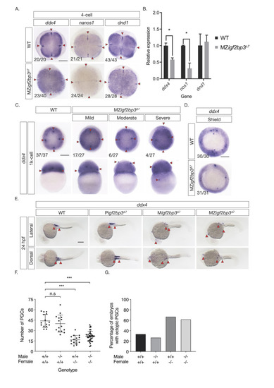
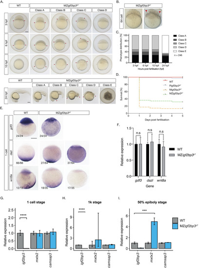
A. Whole mount in-situ hybridisation (WISH) in early embryos shows altered expression of germline markers ddx4, dnd1, and nanos1 in igf2bp3Δ7 mutant embryos. B. qRT-PCR to detect early germplasm markers shows reduced levels of ddx4 and nos1 expression in mutant embryos at the 4-cell stage, whereas expression levels of dnd1 is not significantly different from control wild type embryos. C. Primordial germ cells (PGCs; red arrowheads) in igf2bp3Δ7 embryos are ectopically located at the 1k-cell stage to varying extents ranging from mild or moderate to severe. D. Primordial germ cells are severely reduced or not detected in igf2bp3Δ7 mutants by gastrula stages. E,F. WISH (E) and quantitation (F) of ddx4-positive cells (red arrowheads) shows reduced and ectopic primordial germ cells in 24 hpf maternal igf2bp3Δ7 (Migf2bp3Δ7) and maternal-zygotic igf2bp3Δ7 mutants (MZigf2bp3Δ7) compared to WT siblings and paternal igf2bp3Δ7 (Pigf2bp3Δ7) mutants; p *<0.05, **<0.01, ***< 0.001. G. Loss of maternal igf2bp3 leads to some ectopic primordial germ cells across the trunk (red arrowheads) and occasionally in the hindbrain region. Bar graph shows the number of embryos with ectopic germ cells in WT, Pigf2bp3Δ7, Migf2bp3Δ7, and MZigf2bp3Δ7 mutants. Scale bar in A and C-E, 200 μm. N = 15 embryos each for WT, Pigf2bp3Δ7, and Migf2bp3Δ7 and 39 for MZigf2bp3Δ7 mutants.

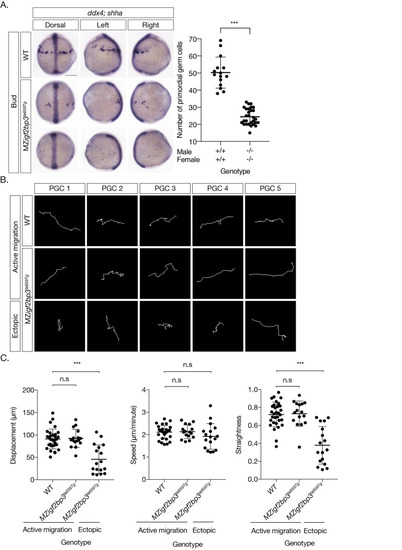
A. WISH to detect ddx4 positive PGCs and shh in the midline at Bud stage. PGCs are reduced (scatter plot, right) and ectopically located in MZigf2bp3la659Tg embryos compared to wild type (WT) embryos. B. Migration tracks of PGCs in bud stage embryos, labelled with GFP-nos1 reporter. MZigf2bp3la659Tg embryos show aberrant PGC migration compared to WT controls. C. PGC track analysis showing displacement, speed and straightness index. Ectopic PGCs in MZigf2bp3la659Tg mutants show significantly reduced total displacement and straightness, although speed of individual PGCs is not altered in mutant embryos compared to WT controls. Scale bars, 200 μm; p*<0.05, **<0.01, ***<0.001; Number of PGCs analysed = 31, 16 and 17 for WT, actively migrating and ectopic MZigf2bp3la659Tg PGCs, respectively.

EXPRESSION / LABELING:
Genes:
Fish:
Anatomical Terms:
Stage: Bud
PHENOTYPE:
Fish:
Observed In:
Stage: Bud

PGC membrane projections show altered behaviour in MZ<italic toggle='yes'>igf2bp3</italic><sup><italic toggle='yes'>la659Tg</italic></sup> mutants.

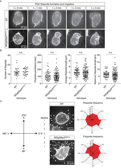
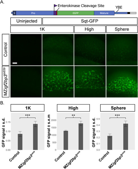
A. Images of cell membranes of PGCs labelled with a Farnesylated-GFP-nos1 live reporter in bud stage mutant or wild type embryos. B. Quantitation of filopodia and behaviour (persistence, length) in WT and MZigf2bp3la659Tg PGCs. C. Projection angle relative to their destination, showing an altered skew in maternal igf2bp3la659Tg PGCs, although the projection parameters, namely numbers, persistence, average and maximum lengths did not appear to be changed. p *<0.05, **<0.01, ***<0.001. N = 27 WT and 26 MZigf2bp3la659Tg PGCs; number of filopodia = 50 WT and 100 mutant filopodia. Number of PGCs analysed for projection frequency = 25 per genotype; Scale bars, 10 μm.

Loss of PGCs in MZ<italic toggle='yes'>igf2bp3</italic><sup><italic toggle='yes'>la659Tg</italic></sup> mutant embryos might arise from cell death.

A. Some PGCs are lost during migration in maternal igf2bp3la659Tg embryos. At bud stage, a subset of PGCs labelled with a GFP-nos1 live reporter display fragmentation during migration. B. Immunofluorescence with an α-Vasa antibody and TUNEL staining showed that compared to wild type embryos, in MZigf2bp3la659Tg embryos a subset of PGCs undergo cell death and fragmentation at the bud stage. Yellow arrow indicates co-localisation of Vasa and TUNEL labelling in a PGC, white arrows indicate Vasa+ fragments; Scale bar, 50 μm.

ZFIN is incorporating published figure images and captions as part of an ongoing project. Figures from some publications have not yet been curated, or are not available for display because of copyright restrictions.

EXPRESSION / LABELING:
Gene:
Fish:
Anatomical Term:
Stage Range: 4-cell to Prim-5
Acknowledgments
This image is the copyrighted work of the attributed author or publisher, and ZFIN has permission only to display this image to its users. Additional permissions should be obtained from the applicable author or publisher of the image. Full text @ PLoS Genet.