FIGURE SUMMARY
Title

A forward genetic screen identifies Dolk as a regulator of startle magnitude through the potassium channel subunit Kv1.1

Authors
Meserve, J.H., Nelson, J.C., Marsden, K.C., Hsu, J., Echeverry, F.A., Jain, R.A., Wolman, M.A., Pereda, A.E., Granato, M.
Source
Full text @ PLoS Genet.

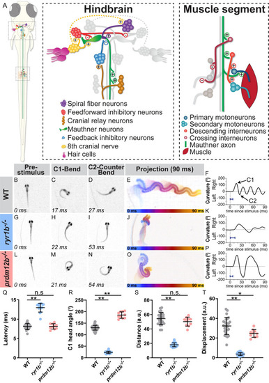
The zebrafish larval startle response is amenable to circuit and genetic analysis.

(A) The acoustic startle response is driven by an action potential from the Mauthner neuron (green), which activates motor neurons in the spinal cord to drive a contralateral body bend. Excitatory and inhibitory neurons in the hindbrain and in the spinal cord impinge upon the Mauthner cells to ensure motor neurons fire on only one side. (B-F) A representative acoustic startle response in a 5 dpf (days post fertilization) wild type larva. An acoustic stimulus is delivered at 0 ms (B), which elicits a rapid turn (C) followed by a counter bend (D) and swimming away (projection of 90 ms response in E, color coded by time). Automated tracking of the curvature of the larva throughout the behavior (F) reveals the response latency (blue bar = latency in F,K,P). (G-K) ryr1b mutants display a weak startle response with reduced bend and counter bend angles (H,I) and reduced displacement (J), largely due to minimal swimming after the counter bend (K). (L-P) prdm12b mutants display an exaggerated startle response with increased bend and counter bend angles (M,N). The duration of each bend is longer than in wild type as well (P). (Q-T) Quantification of response latency (Q; manual measurement); max C1 head angle (R; automated measurement); distance traveled along escape trajectory (S; automated measurement); and displacement from initial head position to final head position (T; automated measurement). Each point is the average response over ten trials for an individual larva. n≥10 larvae, *p = 0.002, **p<0.0001 (one-way ANOVA with Tukey correction for multiple comparisons).

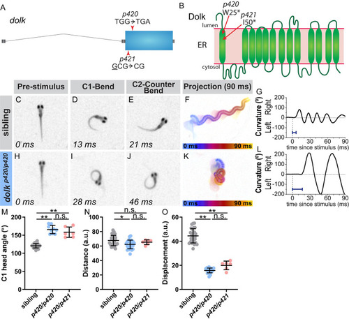
The glycosylation pathway enzyme Dolk regulates the magnitude of the startle response.

(A) Gene structure for dolk with the nonsense mutation from the screen noted (p420). The CRISPR 1 bp deletion allele is also noted (p421). (B) Protein structure for Dolk on the endoplasmic reticulum [48], with the predicted amino acid change from the screen mutation indicated. The deletion in the CRISPR allele causes a frame shift in the sequence that results in a premature stop at amino acid 50. In contrast to siblings (C-G), dolk mutants (H-L) display an exaggerated startle response, with an increased bend and counter bend angle (I,J), resulting in larvae swimming in a “figure eight” (K). Blue bar = latency in G,L. (M-O) Kinematic parameters of the acoustic startle response in dolk siblings and mutants (homozygotes of the screen identified mutation, p420/p420, and transheterozygotes from the screen mutation and CRISPR, p420/p421). Each point represents average of ten trials for an individual fish. n≥6 larvae, **p<0.001, *p = 0.008 (one-way ANOVA with Tukey correction for multiple comparisons).

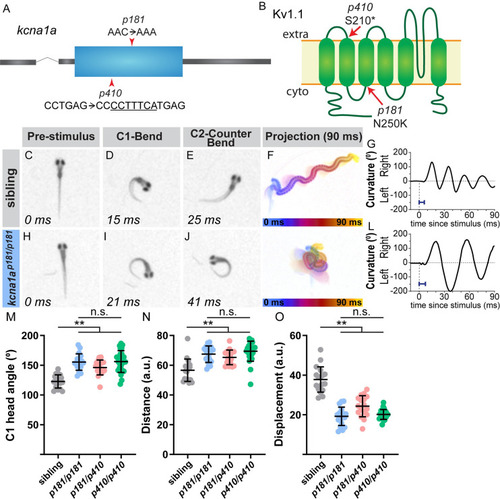
The potassium channel subunit Kv1.1 regulates the magnitude of the startle response.

(A) Gene structure for kcna1a with the missense mutation from the screen noted (p181). The CRISPR 7 bp insertion allele is also noted (p410). (B) Protein structure for Kv1.1, which is encoded by kcna1a, on the plasma membrane, with the predicted amino acid change from the screen allele (p181) indicated. The insertion in the CRISPR allele (p410) causes a frame shift in the sequence that results in a premature stop at amino acid 210. In contrast to siblings (C-G), kcna1a mutants (H-L) display an exaggerated startle response, with an increased bend and counter bend angle (I,J), resulting in fish swimming in a “figure eight” (K). Blue bar = latency in G,L. (M-O) Kinematic parameters of the acoustic startle response in kcna1a sibling and mutants (homozygotes of the screen mutation, p181/p181, transheterozygotes of the screen mutation and CRISPR, p181/p410, and homozygotes of the CRISPR mutation, p410/p410). Each point represents average of ten trials for an individual fish. n≥17 larvae, **p<0.001 (one-way ANOVA with Tukey correction for multiple comparisons).

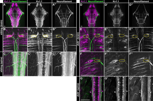
Dolk is required for proper localization of Kv1.1.

(A-C) In 5 dpf dolk sibling larvae, Kv1.1 (magenta:A,B,C; grey:A’,B’,C’) localizes to fiber tracts (α-3A10 antibody stains neurofilament; green:A,B,C; grey:A”,B”,C”) throughout the brain (A; box indicates hindbrain zoom in B) and spinal cord (C). Particularly high accumulation of Kv1.1 is observed at the axon cap (B, yellow dotted circle), where spiral fiber neurons form axo-axonic synapses with the Mauthner cell. (D-G) In dolk mutants, Kv1.1 is localized within somata of neurons throughout the brain and spinal cord and is strongly reduced along axons throughout the hindbrain (D,D’,E,E’). Soma localization is particularly apparent in the large Mauthner neuron, and Kv1.1 is strikingly absent from its axon cap (F, nucleus marked by asterisk, E,F axon cap marked by dotted ovals). Scale bars = 50 μM.

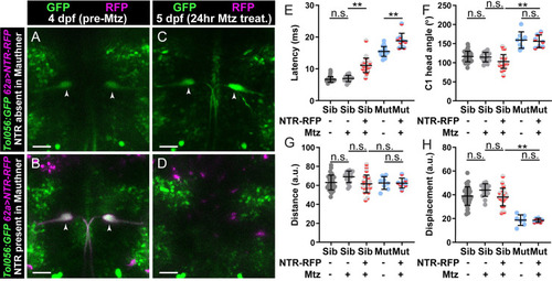
Kv1.1 functions outside the Mauthner command neuron to control swim movement magnitude.

(A-D) kcna1a mutants expressing Tol056 (GFP in Mauthner cells; green), 62a:Gal4 (Mauthner specific) and UAS:NTR-RFP (magenta). 62a>NTR-RFP is mosaic so some fish do not display expression in Mauthner cells (A) while others do (B). Before treatment with metronidazole (Mtz) at 4 dpf, all fish have GFP+ Mauthner cells (A,B). After 24 hr Mtz treatment, Mauthner cells are present in fish without initial NTR-RFP Mauthner expression (C) and absent in fish that displayed initial Mauthner NTR-RFP expression (D). (E-H) Kinematic parameters of the acoustic startle response in kcna1a sibling and mutant larvae with or without NTR-RFP expression in Mauthners and with or without Mtz treatment. Each point is the average response over ten trials for an individual larva. n≥9 larvae, **p<0.0001 (one-way ANOVA with Tukey correction for multiple comparisons). Scale bars = 50 μM.

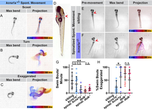
Kv1.1 and Dolk function in the spinal cord to control movement magnitude.

(A-C) Examples of spontaneous swim movements performed by kcna1a mutant larvae. (D) Larvae were transected posterior to the brain to sever the spinal cord. Red arrows indicate the dorsal and ventral points of the cut. Some yolk was left intact to allow mounting of the head in agarose and imaging of the tails, which without the heads would lay on their sides. (E) Example of spinalized sibling and (F) spinalized kcna1a mutant spontaneous movement. (G) The number of spontaneous swim bouts initiated per minute for intact siblings and kcna1a mutants, spinalized kcna1a mutant larvae, intact dolk mutants, and spinalized dolk mutants (H) Percent of spontaneous swim bouts that are abnormal exaggerated movements with high amplitude bends. Each point is an individual fish. n≥6 fish. **p<0.0001, *p = 0.0038 (one-way ANOVA with Tukey correction for multiple comparisons).

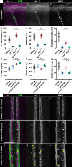
Kv1.1 is expressed in motor neurons and V2a interneurons.

(A) Kv1.1 (magenta:A; grey:A’) localizes to motor axons expressing hb9:GFP (green:A; grey:A”). (B-G) Kinematic parameters of the acoustic startle response in (B-D) sibling, ryr1b single mutants, dolk single mutants, and ryr1b-/-;dolk-/- double mutants, and (E-G) sibling, kcna1a single mutants, prdm12b single mutants, and kcna1a-/-;prdm12b-/- double mutants. Each point represents average of ten trials for an individual fish. n≥6 larvae, *p<0.04 **p<0.0001 (one-way ANOVA with Tukey correction for multiple comparisons). (H-I) Kv1.1 (magenta:H,I; grey:H’,I’) localizes to (H) V2a interneuron axons expressing chx10::GFP (green:H,I; grey:H”,I”) in the ventral spinal cord but not (I) V2a interneuron soma. (J) In dolk mutants, Kv1.1 (magenta:J; grey:J’) localizes to V2a interneuron soma expressing chx10::GFP (green:J; grey:J”). Scale bars = 50 μM.

Acknowledgments
This image is the copyrighted work of the attributed author or publisher, and ZFIN has permission only to display this image to its users. Additional permissions should be obtained from the applicable author or publisher of the image. Full text @ PLoS Genet.