IMAGE

Fig 6

ID
ZDB-IMAGE-210623-21
Source
Figures for Meserve et al., 2021
Image
Figure Caption

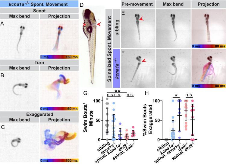
Fig 6 Kv1.1 and Dolk function in the spinal cord to control movement magnitude.

(A-C) Examples of spontaneous swim movements performed by kcna1a mutant larvae. (D) Larvae were transected posterior to the brain to sever the spinal cord. Red arrows indicate the dorsal and ventral points of the cut. Some yolk was left intact to allow mounting of the head in agarose and imaging of the tails, which without the heads would lay on their sides. (E) Example of spinalized sibling and (F) spinalized kcna1a mutant spontaneous movement. (G) The number of spontaneous swim bouts initiated per minute for intact siblings and kcna1a mutants, spinalized kcna1a mutant larvae, intact dolk mutants, and spinalized dolk mutants (H) Percent of spontaneous swim bouts that are abnormal exaggerated movements with high amplitude bends. Each point is an individual fish. n≥6 fish. **p<0.0001, *p = 0.0038 (one-way ANOVA with Tukey correction for multiple comparisons).

Figure Data
Acknowledgments
This image is the copyrighted work of the attributed author or publisher, and ZFIN has permission only to display this image to its users. Additional permissions should be obtained from the applicable author or publisher of the image. Full text @ PLoS Genet.