
- Title
-
IL-1R1-Dependent Signals Improve Control of Cytosolic Virulent Mycobacteria In Vivo
- Authors
- van der Niet, S., van Zon, M., de Punder, K., Grootemaat, A., Rutten, S., Moorlag, S.J.C.F.M., Houben, D., van der Sar, A.M., Bitter, W., Brosch, R., Hernandez Pando, R., Pena, M.T., Peters, P.J., Reits, E.A., Mayer-Barber, K.D., van der Wel, N.N.
- Source
- Full text @ mSphere

The pH of the phagolysosome does not affect the cytosolic translocation of |

The cytosolic localization of M. marinum in zebrafish is abundant when the adaptive immune system is not yet developed. Embryo and adult zebrafish infected with M. marinum were analyzed using TEM. (A) Cytosolic M. marinum in zebrafish embryo tissue. (B) Phagosomal M. marinum ΔESX-5 bacteria in adult zebrafish tissue. (A′ and B′) Schematic representations of panels A and B, with black dots indicating actin immunogold labeling, orange lines indicating M. marinum, blue lines indicating phagosomal and host membranes, and green lines indicating mitochondria. (C) Quantification of the percentage of cytosolic M. marinum bacteria at embryo day 9 and in the spleen of adult zebrafish at day 11 (see also Fig. S2B in the supplemental material). Error bars indicate standard deviations between 3 different zebrafish embryos and 3 adult fish, and n represents the total number of bacteria counted. |
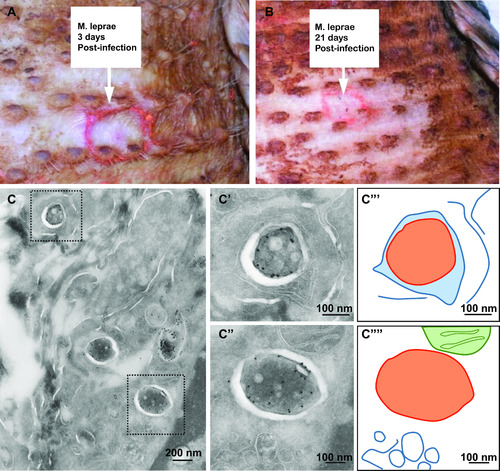
Restrained cytosolic |

Restrained cytosolic localization of |

Early cytosolic localization of |

M. tuberculosis preferentially localizes to the cytosol in Il1r1−/− mice. (A and B) Fluorescence microscopy of 200-nm sections stained with DAPI (4′,6-diamidino-2-phenylindole) for nuclei (white) and anti-cell wall protein to detect M. tuberculosis (green) in granulomas in lung tissues of WT B6 mice (A) and Il1r1−/− mice (B) infected with M. tuberculosis for 28 days. (C) Immunogold labeling using LAMP1 to indicate lysosomal membranes in Il1r1−/− mice imaged using TEM. (C′) Schematic representation of panel C. M. tuberculosis is depicted in orange, host membranes are in blue, and the host nucleus is in pink. (D) Quantification of the localization of M. tuberculosis in B6 and Il1r1−/− lungs, here presenting cytosolic localization (see also Fig. S2D in the supplemental material). The analysis included 897 (WT) or 618 (Il1r1−/−) bacteria, and error bars indicate standard deviations based on in multiple granulomas of 2 WT B6 and 2 Il1r1−/− mice. KO, knockout. |