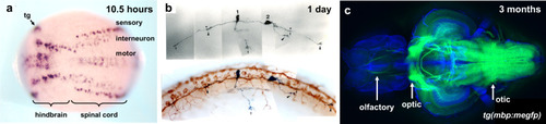
Development of the central nervous system in zebrafish.
a Detection of early neuronal precursor cells by whole-mount in situ hybridization with a pan-neuronal marker, huC, at the neural plate stage (10.5 h after fertilization). Unpublished data. b Immunostaining of axonal growth in the spinal cord of one-day-old zebrafish. Double-staining with anti-gicerin antibody and anti-HNK-1 antibody28. c Confocal image of myelin structure in an isolated adult zebrafish brain visualized by mbp promoter-driven membrane-tagged GFP, Tg(mbp:mEGFP). Arrows indicate the olfactory, optic, and otic nerves. Unpublished data.
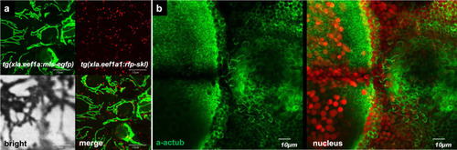
Subcellular organelles in the developing zebrafish embryos.
a Using transgenic zebrafish lines 5 dpf, mitochondria, Tg(Xla.Eef1a:MLS-EGFP), and peroxisomes, Tg(Xla. Eef1a:RFP-SKL), in the skin of the developing larva are visualized. b Motile cilia (green) in the hindbrain 4th ventricle are visualized with anti-acetylated tubulin antibody, and nuclei are shown in red. Unpublished data.
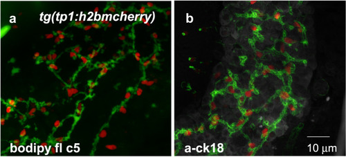
Bile duct formation in the developing zebrafish liver 6 dpf.
a, b Using a transgenic zebrafish line, Tg(Tp1:H2BmCherry), biliary epithelial cell nuclei are labeled red. The bile duct in the developing liver is visualized using the BODIPY FL-C5 dye (a) or the anti-cytokeratin 18 antibody (b). Unpublished data.
Acknowledgments
This image is the copyrighted work of the attributed author or publisher, and
ZFIN has permission only to display this image to its users.
Additional permissions should be obtained from the applicable author or publisher of the image.
Full text @ Exp. Mol. Med.
Your Input Welcome
Thank you for submitting comments. Your input has been emailed to ZFIN curators who may contact you if
additional information is required.
Oops. Something went wrong. Please try again later.