FIGURE SUMMARY
Title

Phosphorylation of seryl-tRNA synthetase by ATM/ATR is essential for hypoxia-induced angiogenesis

Authors
Shi, Y., Liu, Z., Zhang, Q., Vallee, I., Mo, Z., Kishi, S., Yang, X.L.
Source
Full text @ PLoS Biol.

SerRS is involved in the hypoxia response to induce VEGFA expression through phosphorylation by ATM and ATR kinases at S101 and S241 residues.

(A, B) qRT-PCR analysis of VEGFA expression in HEK293 cells transfected with sh-SerRS, sh-GlyRS, or nonspecific sh-Control under hypoxia or normoxia conditions. VEGFA levels (A) and relative induction of VEGFA under hypoxia (B) were plotted as means ± SEM (n = 4, biological replicates, Student t test, **p < 0.01, ****p < 0.0001). (C) Sequence alignment of SerRS proteins from vertebrates and non-vertebrates flanking serine 101 and serine 241 (highlighted in red) sites numbered according to the human sequence. The conserved ATM/ATR substrate motif residues are underlined. (D) 32P-labeling to confirm phosphorylation of SerRS, but not GlyRS, in vitro using recombinant tRNA synthetases. Double-stranded DNA oligonucleotides are used to mimic DNA damage to activate ATM/ATR from the nuclear extract of HEK293 cells. (E) Western blot analysis to confirm recombinant SerRS, but not GlyRS and TyrRS, is phosphorylated by ATM/ATR by using a specific antibody against phosphor-ATM/ATR substrate (p-S*Q). The purified His6-tagged aaRS proteins are recognized by anti-His6-tag antibody. Activation of ATM and ATR was confirmed by autophosphorylation of ATM (at S1981) and phosphorylation of RPA32 (at S33), respectively. (F, G) IP and western blot analysis to confirm hypoxia-induced SerRS phosphorylation in HEK293 (F) and HUVEC (G) cells. IP was performed with anti-SerRS antibody from mouse and rabbit for HEK293 (F) and HUVEC (G) cells, respectively. Phosphorylated SerRS was detected with the antibody against phosphor-ATM/ATR substrate (p-S*Q) from rabbit. Activation of ATM and ATR was confirmed by autophosphorylation of ATM (at S1981) and phosphorylation of CHK1 (at S345) and P53 (at S15). (H) Hypoxia-induced phosphorylation of SerRS in HEK293 cells is reduced in SerRSAA compared with SerRSWT. (I) Hypoxia-induced phosphorylation of SerRS was decreased when ATM and ATR were knocked down separately or together by siRNAs (si-ATM, si-ATR). At least 2 biological replicates were performed for western blot experiments with consistent results. Representative images were shown. See S1 Data for quantitative data and statistical analysis. See S2 Data for original, uncropped images supporting blots and gel results. aaRS, aminoacyl-tRNA synthetase; ATM, ataxia telangiectasia mutated; ATR, ataxia telangiectasia mutated and RAD3-related; GlyRS, glycyl-tRNA synthetase; HUVEC, human umbilical vein endothelial cell; IP, immunoprecipitation; qRT-PCR, real-time quantitative reverse transcription PCR; SerRS, seryl-tRNA synthetase; SerRSAA, S101A/S241A; SerRSWT, wild-type SerRS; sh-Control, control shRNA; sh-GlyRS, shRNAs targeting GlyRS; sh-SerRS, shRNAs targeting SerRS; siRNA, small interfering RNA; TyrRS, tyrosyl-tRNA synthetase; VEGFA, vascular endothelial growth factor A.

SerRS phosphorylation at S101 and S241 inhibits its function in repressing VEGFA expression in cellulo and in vivo.

(A) qRT-PCR analysis of VEGFA expression in HEK293 cells transfected with Flag-tagged SerRSWT or mutants with SerRSAA and SerRSDD substitutions. The exogenous expressions of the SerRS proteins were confirmed by immunoblotting against the Flag tag. VEGFA expression levels are plotted as means ± SEM (n = 3, biological replicates, ****p < 0.0001, Student t test). (B) qRT-PCR analysis of vegfaa transcription in zebrafish injected with SerRS-MO and with co-injection of SerRS-MO and SerRSWT, SerRSAA, or SerRSDD mRNA. Data plotted as means ± SEM (n = 3–4, biological replicates, **p < 0.01, ***p < 0.001, Student t test). The Coomassie blue staining was used to show equal total protein loading. See S1 Data for quantitative data and statistical analysis. See S2 Data for original, uncropped images supporting blots and gel results. qRT-PCR, real-time quantitative reverse transcription PCR; SerRS, seryl-tRNA synthetase; SerRSAA, S101A/S241A; SerRSDD, S101D/S241D; SerRS-MO, antisense morpholino against SerRS; SerRSWT, wild-type SerRS; VEGFA, vascular endothelial growth factor A.

EXPRESSION / LABELING:
Gene:
Fish:
Knockdown Reagent:
Anatomical Term:
Stage: Protruding-mouth
PHENOTYPE:
Fish:
Knockdown Reagent:
Observed In:
Stage: Protruding-mouth

ATM/ATR-caused SerRS phosphorylation attenuates its binding to <italic>VEGFA</italic> promoter, which allows c-Myc and HIF-1 to activate <italic>VEGFA</italic> transcription.

(A) EMSA to measure the binding of SerRSWT, SerRSAA, or SerRSDD with 32P-labeled DNA fragments (27 bp) corresponding to the SerRS binding site on human VEGFA promoter. (B) Chromatin-immunoprecipitated DNA by SerRS antibody was quantified by qPCR (ChIP-qPCR) to measure the binding of SerRSWT, SerRSAA, or SerRSDD on VEGFA promoter in HEK293 cells. The data were calculated as percentage of input DNA and then normalized against the SerRSWT group (means ± SEM, n = 2, biological replicates, ****p < 0.0001, Student t test). (C) Structure model of SerRS dimer binding to VEGFA promoter DNA showing that the phosphorylation sites S101 and S241 are located near the predicted SerRS-DNA binding sites. (D, E) ChIP-qPCR to follow the binding of endogenous SerRS, c-Myc, and HIF-1α on VEGFA promoter during hypoxia in HUVEC cells (D) and HEK293 cells (E) (means ± SEM, n = 3, biological replicates, **p < 0.01, Student t test). (F) ChIP-qPCR to measure the binding of SerRS, c-Myc, and HIF-1α on VEGFA promoter in HEK293 cells stably transfected with SerRSWT, SerRSAA, or SerRSDD (tag-free) under normoxia and hypoxia (12 h) conditions (means ± SEM, n = 3, biological replicates, **p < 0.01, ***p < 0.001, ****p < 0.0001, Student t test). The expression level of the exogenous SerRS proteins (WT, AA, and DD) is similar (as shown in Fig 4A and S5 Fig). (G) qRT-PCR to measure VEGFA expression in HEK293 cells transfected with SerRSWT or SerRSAA under normoxia and hypoxia conditions (means ± SEM, n = 4, biological replicates, *p < 0.05, **p < 0.01, n.s., Student t test). (H) qRT-PCR to measure the effects of specific ATM inhibitor KU-55933 and specific ATR inhibitor VE-821 on hypoxia-induced VEGFA expression in HEK293 cells (means ± SEM, n = 2, biological replicates, *p < 0.05, ****p < 0.0001, Student t test). (I) ChIP-qPCR to measure the binding of SerRS to VEGFA promoter in HEK293 cells stably transfected with SerRSWT, SerRSAA, or SerRSDD under normoxia or hypoxia, in the presence or absence of VE-821 (means ± SEM, n = 3, biological replicates, **p < 0.01, ***p < 0.001, Student t test). See S1 Data for quantitative data and statistical analysis. See S2 Data for original, uncropped images supporting blots and gel results. ATM, ataxia telangiectasia mutated; ATR, ataxia telangiectasia mutated and RAD3-related; ChIP-qPCR, chromatin immunoprecipitation–quantitative real-time PCR; EMSA, electrophoresis mobility shift assay; HIF-1; hypoxia-inducible factor 1; HUVEC, human umbilical vein endothelial cell; n.s., not significant; qRT-PCR, real-time quantitative reverse transcription PCR; SerRS, seryl-tRNA synthetase; SerRSAA, S101A/S241A; SerRSDD, S101D/S241D; SerRSWT, wild-type SerRS; VEGFA, vascular endothelial growth factor A.

SerRS phosphorylation is required for activating VEGFA-mediated tube formation under hypoxia.

(A, B) VEGFA levels in HEK293 (A) and HUVEC (B) cells stably transfected with SerRSWT, SerRSAA, or SerRSDD under normoxia or hypoxia conditions were measured by ELISA and normalized to total proteins in cell lysates. (means ± SEM, n = 2 biological replicates, *p < 0.05, **p < 0.05, n.s., Student t test). The SerRS protein levels were shown by western blot below the bar graph. (C, D) Endothelial cell tube formation assay (C) and quantification (D) using HUVEC cells treated with indicated conditional medium from wild-type or mutant SerRS stably transfected donor HUVEC cells cultured under normoxia or hypoxia conditions. The cartoon describes the experimental setup. The total length of tubules was measured using Image J software (means ± SEM, n = 3, biological replicates, *p < 0.05, **p < 0.01, ***p < 0.001, n.s., Student t test). SeeS1 Data for quantitative data and statistical analysis. See S2 Data for original, uncropped images supporting blots and gel results. HUVEC, human umbilical vein endothelial cell; n.s., not significant; SerRS, seryl-tRNA synthetase; SerRSAA, S101A/S241A; SerRSDD, S101D/S241D; SerRSWT, wild-type SerRS; VEGFA, vascular endothelial growth factor A.

SerRS<sup>AA</sup> exhibits stronger activity than SerRS<sup>WT</sup> in suppressing angiogenesis in mice.

(A) Western blot to measure the expression of SerRS proteins in mouse 3B11 endothelial cells stably transfected with tag-free mouse SerRSWT or mutants (SerRSAA and SerRSDD). The SerRS levels are quantified by the density of the bands and indicated relative to the level of the endogenous SerRS (Vector). (B, C) Matrigel plug angiogenesis assay performed with stably transfected 3B11 cells in C3H/HeJ mice. Matrigel plugs (dash lines enclosed regions) excised 14 days after implantation were analyzed by immunofluorescence for Vegfa (B) and quantified (C) (means ± SEM, n = 6 individual plugs per group, *p < 0.05, **p < 0.01, ***p < 0.001, n.s., Student t test). See S1 Data for quantitative data and statistical analysis. See S2 Data for original, uncropped images supporting blots and gel results. n.s., not significant; SerRS, seryl-tRNA synthetase; SerRSAA, S101A/S241A; SerRSDD, S101D/S241D; SerRSWT, wild-type SerRS; Vegfa, vascular endothelial growth factor A

SerRS<sup>AA</sup> exhibits stronger activity than SerRS<sup>WT</sup> in inhibiting tumor angiogenesis and tumor growth in mice.

(AC) Tumor xenografts formed by the original (control) or engineered MDA-MB-231 cells were analyzed by monitoring the growth curves (A) and comparing the size of the xenografts at end point (22 days post implantation) (B) with quantifications (C). (DF) The dissected MDA-MB-231 breast cancer xenografts were analyzed by western blots for SerRS, HIF-1α, VEGFA, Cdh5, and β-actin protein levels (D), and the levels were quantified for VEGFA (E) and Cdh5 (F). (GJ) Tumor angiogenesis in MDA-MB-231 xenografts was analyzed by immunofluorescent staining of VEGFA (G) and Cdh5 (I) and quantified by measuring the fluorescent signal intensities (H, J). (K, L) IP and western blot analysis to confirm SerRS phosphorylation in tumor xenografts. IP was performed with anti-SerRS antibody from rabbit before phosphorylated SerRS was detected with a specific antibody against p-S*Q (K) and quantified by measuring the band intensities (L). Data are shown as means ± SEM, n = 5 mice, *p < 0.05, **p < 0.01, ***p < 0.001, n.s., Student t test). See S1 Data for quantitative data and statistical analysis. See S2 Data for original, uncropped images supporting blots and gel results. HIF-1α; hypoxia-inducible factor 1α; IP, immunoprecipitation; n.s., not significant; SerRS, seryl-tRNA synthetase; SerRSAA, S101A/S241A; SerRSDD, S101D/S241D; SerRSWT, wild-type SerRS; VEGFA, vascular endothelial growth factor A.

Schematic diagram to illustrate the impact of SerRS in hypoxia-induced angiogenesis.

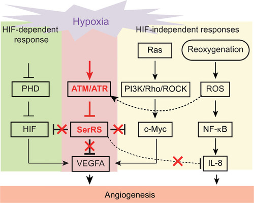
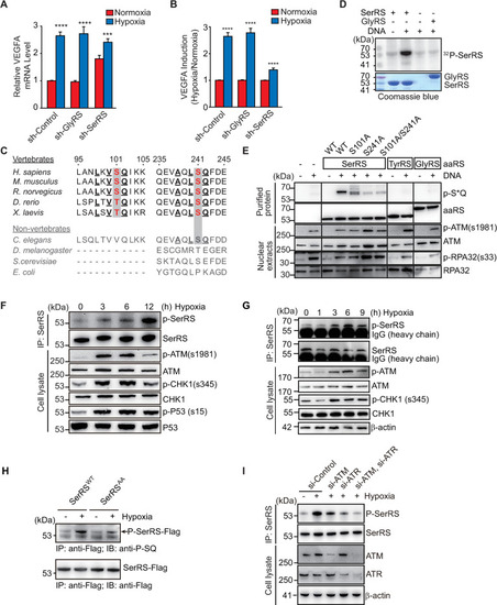
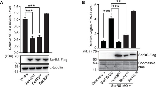
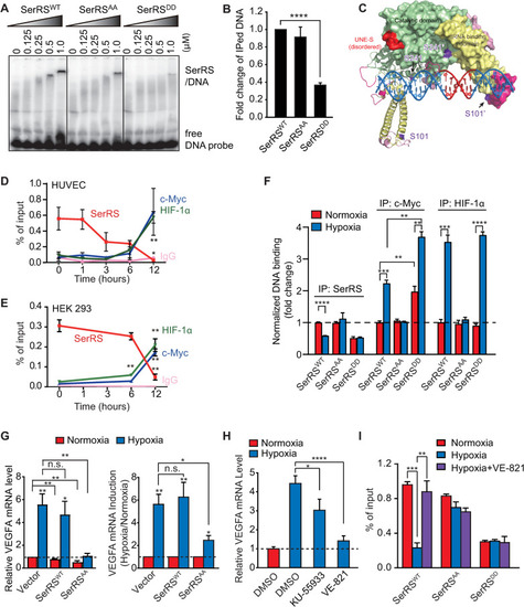
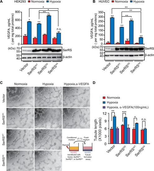
Multiple pathways are activated under hypoxia to induce angiogenesis. SerRS has the capacity to inhibit these pathways regardless of whether they are dependent or independent of HIF. This capacity, however, is silenced by ATM/ATR, which is also activated by hypoxia, highlighting the central role of SerRS in regulating angiogenesis. ATM, ataxia telangiectasia mutated; ATR, ataxia telangiectasia mutated and RAD3-related; HIF; hypoxia-inducible factor; SerRS, seryl-tRNA synthetase.

ZFIN is incorporating published figure images and captions as part of an ongoing project. Figures from some publications have not yet been curated, or are not available for display because of copyright restrictions.

PHENOTYPE:
Fish:
Knockdown Reagent:
Observed In:
Stage: Protruding-mouth
Acknowledgments
This image is the copyrighted work of the attributed author or publisher, and ZFIN has permission only to display this image to its users. Additional permissions should be obtained from the applicable author or publisher of the image. Full text @ PLoS Biol.