FIGURE SUMMARY
Title

Pharyngeal pouches provide a niche microenvironment for arch artery progenitor specification

Authors
Mao, A., Zhang, M., Li, L., Liu, J., Ning, G., Cao, Y., Wang, Q.
Source
Full text @ Development

Pharyngeal mesoderm contains two vascular progenitor subpopulations. (A) In situ hybridization analysis of the expression of ZsYellow and nkx2.5. Numbers indicate PAA clusters. Lateral views with anterior on the left. (B,C) The expression patterns of the PAA progenitor marker nkx2.5 (B), and angioblast markers scl and etv2 (C) in Tg(nkx2.5:ZsYellow) embryos were detected by fluorescent in situ hybridization. Embryos were first subjected to fluorescent in situ hybridization and then to immunostaining with anti-ZsYellow antibody. Scale bar: 20 μm in B; 50 μm in C. (D) Colocalization analysis of nkx2.5 and etv2 by double fluorescent in situ hybridization. Scale bar: 20 μm. (E-G) The Kaede proteins in PAA cluster 3 and the subsequent posterior pharyngeal mesoderm on the right side of the Tg(nkx2.5:kaede) embryos were photoconverted at 22 hpf (E). Cells on the left side of the pharyngeal mesoderm remained unphotoconverted as an internal control (inset). Embryos were subsequently imaged in the green and red channels at 36 hpf (F) and 60 hpf (G). Scale bars: 50 μm. (H-K) Localized photoconversion of PAA cluster 5 (H) or Kaede+ cells between cluster 4 and 5 (J) at 36 hpf. Red, green and merged images of the same embryos at 54 hpf are shown in I and K. Scale bars: 50 μm. The photoconversion experiments in E-K were repeated three times independently, and three to five embryos per group were used each time. All the embryos analyzed showed similar distributions of photoconverted cells. The Kaede+ cells in PAA cluster 5 and between PAA clusters 4 and 5 gave rise to PAA5 and ventral aorta, respectively, indicating that the pharyngeal mesoderm contains two vascular progenitor subpopulations.

Ablation of pouch endoderm impairs PAA progenitor specification. (A) Confocal images of live Tg(nkx2.5:ZsYellow;nkx2.3:mCherry) embryos. The formation of mCherry+ pharyngeal pouches (red) coincides with the emergence of ZsYellow+ PAA clusters (green). Pharyngeal pouches and PAA clusters or sprouts are numbered with yellow or green numbers, respectively. Scale bar: 50 μm. (B) Confocal images of live Tg(nkx2.5:ZsYellow) embryos showed that injection of 8 ng sox32 MO resulted in complete absence of PAA sprouts and PAA tubular structures. The ratios of affected embryos are indicated. Scale bar: 50 μm. (C,D) Embryos were exposed to 10 mM MTZ from bud stage to 38 hpf, and then harvested at the indicated developmental stages for in situ hybridization (C) or in vivo confocal imaging (D). Numbers in C indicate PAA3-PAA6. Scale bar: 50 μm. (E,E′) The scl, etv2 and tie1 transcripts were evaluated by in situ hybridization in Tg(nkx2.3:KalTA4-p2a-mCherry;UAS:NTR-mCherry) embryos treated with DMSO or 10 mM MTZ (E). The average numbers of scl+, etv2+ and tie1+ PAA angioblast clusters were quantified from three independent experiments and the group values are expressed as mean±s.d. (E′). Student's t-test, ***P<0.001. (F) Expression analysis of nkx2.5 in pouch endoderm-depleted embryos. Black arrowheads represent the expression of nkx2.5 in the developing heart.

Pouch endoderm is necessary for BMP signal activation in the presumptive PAA progenitors. (A) The bmp2a, bmp2b, bmp4 and bmp5 transcripts were evaluated by in situ hybridization in Tg(nkx2.3:KalTA4-p2a-mCherry;UAS:NTR-mCherry) embryos treated with DMSO or 10 mM MTZ. (B) BMP signal was dynamically activated in the forming PAA clusters. Tg(nkx2.5:ZsYellow) embryos were harvested at indicated stages and subjected to immunostaining for p-Smad1/5/8 (red) and ZsYellow (green). The white dotted lines outline the PAA progenitor clusters and the purple dotted lines indicate the PAA sprouts composed of migrating angioblasts. Scale bar: 20 μm. (C) Tg(BRE:EGFP;nkx2.5:ZsYellow) embryos were immunostained for GFP (green) and ZsYellow (red) to visualize BMP-responsive cells and PAA clusters. Scale bar: 50 μm. (D) p-Smad1/5/8 levels were greatly decreased in pouch endoderm-depleted embryos. Tg(nkx2.5:ZsYellow;nkx2.3:KalTA4-p2a-mCherry;UAS:NTR-mCherry) embryos were treated with DMSO or 10 mM MTZ from bud stage to 36 hpf, and then stained for p-Smad1/5/8 (red) and ZsYellow (green). Scale bar: 50 μm. (E) Live confocal images of Tg(BRE:EGFP;nkx2.3:KalTA4-p2a-mCherry;UAS:NTR-mCherry) embryos treated with DMSO or 10 mM MTZ from bud stage to 36 hpf. Scale bar: 50 μm. (F) Tg(sox17:GFP) embryos were injected with 40 pg nkx2.3:noggin3-mCherry plasmids and 200 pg Tol2 transposase mRNA at the one-cell stage, and then harvested for in vivo confocal imaging to visualize pharyngeal pouches (green) and the expression of Noggin3-mCherry fusion proteins (red). Scale bar: 50 μm. (G) Pouch-derived Noggin3 significantly decreased the BMP signal activity in the pharyngeal region. Tg(nkx2.5:ZsYellow;BRE:EGFP) embryos were injected with 40 pg nkx2.3:noggin3-mCherry plasmids and 200 pg Tol2 transposase mRNA at the one-cell stage, and then embryos with abundant mCherry fluorescence in the pouches were selected at 36 hpf for immunostaining. Scale bar: 50 μm.

Inhibition of BMP signaling impairs PAA progenitor specification. (A,A′) Transcripts of scl and etv2 were evaluated by in situ hybridization in DMSO- or DMH1-treated embryos. Embryos were treated with DMSO or 10 μM DMH1 from 24 to 38 hpf. Numbers indicate PAA angioblast clusters. The average numbers of scl+ and etv2+ PAA angioblast clusters are quantified in A′ based on three independent experiments and the group values are expressed as mean±s.d. Student's t-test, ***P<0.001. (B,C) Wild-type embryos were treated with DMSO or with 10 μM DMH1 within the indicated time windows. Subsequently, these embryos were harvested for in situ hybridization. The numbers indicate the PAAs (B) or PAA progenitor clusters (C). (D-E′) Ectopic induction of caBmpr1b rescues the defects of PAA progenitor specification and angioblast differentiation in the pouch-depleted embryos. Tg(nkx2.3:KalTA4-p2a-mCherry;UAS:NTR-mCherry;hsp70l:caBmpr1b-GFP) embryos were treated with 10 mM MTZ from bud stage, heat shocked at 24 hpf for 20 min, and then harvested at the indicated stages for in situ hybridizations with nkx2.5 (D) and etv2 (E) probes. The average numbers of etv2+ PAA angioblast clusters were quantified from three independent experiments (E′). Significance of differences compared with MTZ treatment group were analyzed with Student's t-test (*P<0.05; ***P<0.001).

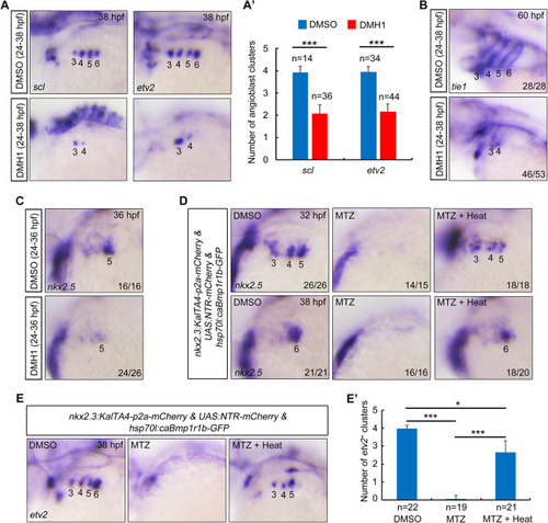
Inhibition of BMP signaling after PAA progenitor specification induces no detectable defects of PAA morphogenesis. (A) Live confocal images of Tg(nkx2.5:ZsYellow;gata1:DsRed) embryos with DMSO or 10 μM DMH1 treatment for different durations. White arrowheads indicate the new sprouting clusters after removal of DMH1 at 38 hpf. Scale bar: 50 μm. (B) Expression analysis of nkx2.5 by in situ hybridization in embryos subjected to different treatments. The dotted lines represent the area of expression of nkx2.5. Black arrowhead indicates the newly emerged expression of nkx2.5 after removal of DMH1.

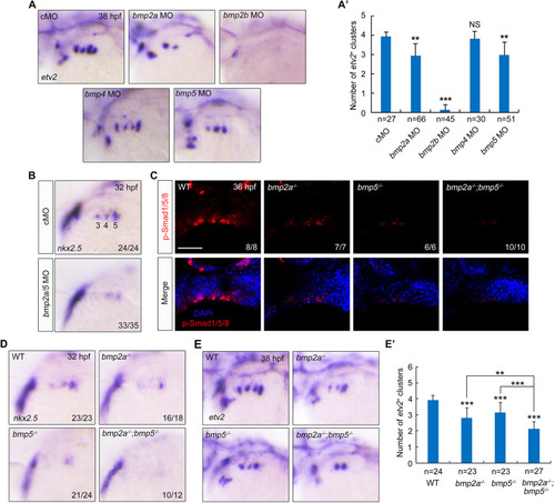
BMP2a together with BMP5 functions in PAA progenitor specification. (A,A′) The expression of etv2 in embryos injected with indicated MO was analyzed by in situ hybridization (A). Injection doses: bmp2a MO, 2 ng; bmp2b MO, 0.3 ng; bmp4 MO, 2 ng; bmp5 MO, 4 ng. The average numbers of etv2+ PAA angioblast clusters were quantified from three independent experiments, and the group values are expressed as mean±s.d. (A′). Student's t-test (**P<0.01, ***P<0.001; NS, no significant difference). (B) Knockdown of bmp2a and bmp5 disrupted the specification of PAA progenitors. Wild-type embryos were injected with bmp2a and bmp5 MOs at the one-cell stage. The resulting embryos were harvested for in situ hybridization. (C) Wild-type and indicated mutant embryos were harvested at 36 hpf for immunofluorescence assay with anti-p-Smas1/5/8 antibody. Nuclei were counterstained with DAPI. There is a distinct decrease of p-Smad1/5/8 in the bmp2a−/−;bmp5−/− double mutants. Scale bar: 50 μm. (D-E′) Expression analysis of nkx2.5 (D) and etv2 (E) in bmp2a−/− or bmp5−/− embryos and bmp2a−/−;bmp5−/− double mutants by in situ hybridization. The average numbers of etv2+ angioblast clusters were quantified from three independent experiments and the group values were expressed as mean±s.d. (E′). Student's t-test was used to determine the significance of differences between wild-type animals and each mutant, and one-way ANOVA test was performed to analyze the statistical differences between bmp2a−/−;bmp5−/− double mutants and bmp2a−/− or bmp5−/− embryos. **P<0.01, ***P<0.001.

npas4l is expressed in both PAA progenitors and angioblasts. (A,B) Analysis of the expression patterns of nkx2.5 and npas4l in the pharynx at the indicated stages by in situ hybridization. The asterisks indicate the expression of npas4l in the lateral dorsal aortae. (C) Double in situ hybridization of npas4l (red) and nkx2.5 (blue) expression at 28 and 36 hpf. (D,E) Expression analysis of nkx2.5 (D) and npas4l (E) in embryos injected with 3 ng nkx2.5 MO. The expression of nkx2.5 but not npas4l was clearly increased in nkx2.5 morphants.

npas4l plays a pivotal role in the specification of PAA progenitors. (A) Live confocal images of npas4l/− mutants and their wild-type and heterozygous siblings in Tg(nkx2.5:ZsYellow) background. Scale bar: 50 μm. The npas4l gene is also called cloche. (B,C) The expression patterns of nkx2.5 (B) and etv2 (C) were analyzed in npas4l−/− mutants and their siblings by in situ hybridization. (D,E) The expression of myod1 (D) and actn3b (E) in the pharynx were analyzed in npas4l−/− mutants and their siblings in Tg(nkx2.5:ZsYellow) background. The embryos were first subjected to fluorescent in situ hybridization with myod1 or actn3b probe, and then stained with anti-ZsYellow antibody. (F) Tg(nkx2.3:KalTA4-p2a-mCherry;UAS:NTR-mCherry) embryos were treated with 10 mM MTZ from bud stage and wild-type embryos were treated with 10 μM DMH1 from 24 hpf or injected with 2 ng bmp2a MO together with 4 ng bmp5 MO at the one-cell stage. The resulting embryos were harvested for in situ hybridization with npas4l probe at 38 hpf. (G,G′) Expression analysis of npas4l in bmp2a−/− or bmp5−/− single mutants and bmp2a−/−;bmp5−/− double mutant embryos (G). The average numbers of npas4l+ clusters were quantified from three independent experiments and the group values are expressed as mean±s.d. (G′). Student's t-test was used to determine the significance of differences between wild-type animals and indicated mutants, and one-way ANOVA test was performed to analyze the statistical differences between bmp2a−/−;bmp5−/− embryos and bmp2a−/− or bmp5−/− single mutants. **P<0.01, ***P<0.001. (H) Working model depicting that the specification of PAA progenitors from pharyngeal mesoderm is dependent of the activation of BMP signaling by bmp2a and bmp5 expressed in pouch endoderm.

Acknowledgments
This image is the copyrighted work of the attributed author or publisher, and ZFIN has permission only to display this image to its users. Additional permissions should be obtained from the applicable author or publisher of the image. Full text @ Development