IMAGE

Fig. 3.

ID
ZDB-IMAGE-210215-43
Source
Figures for Mao et al., 2020
Image
Figure Caption

Fig. 3.

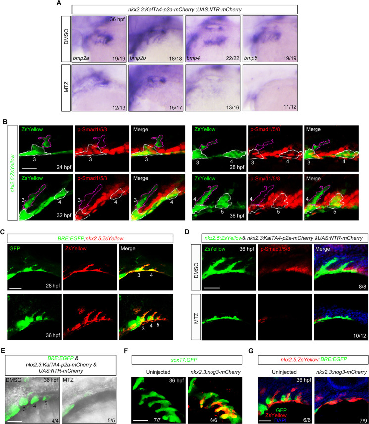
Pouch endoderm is necessary for BMP signal activation in the presumptive PAA progenitors. (A) The bmp2a, bmp2b, bmp4 and bmp5 transcripts were evaluated by in situ hybridization in Tg(nkx2.3:KalTA4-p2a-mCherry;UAS:NTR-mCherry) embryos treated with DMSO or 10 mM MTZ. (B) BMP signal was dynamically activated in the forming PAA clusters. Tg(nkx2.5:ZsYellow) embryos were harvested at indicated stages and subjected to immunostaining for p-Smad1/5/8 (red) and ZsYellow (green). The white dotted lines outline the PAA progenitor clusters and the purple dotted lines indicate the PAA sprouts composed of migrating angioblasts. Scale bar: 20 μm. (C) Tg(BRE:EGFP;nkx2.5:ZsYellow) embryos were immunostained for GFP (green) and ZsYellow (red) to visualize BMP-responsive cells and PAA clusters. Scale bar: 50 μm. (D) p-Smad1/5/8 levels were greatly decreased in pouch endoderm-depleted embryos. Tg(nkx2.5:ZsYellow;nkx2.3:KalTA4-p2a-mCherry;UAS:NTR-mCherry) embryos were treated with DMSO or 10 mM MTZ from bud stage to 36 hpf, and then stained for p-Smad1/5/8 (red) and ZsYellow (green). Scale bar: 50 μm. (E) Live confocal images of Tg(BRE:EGFP;nkx2.3:KalTA4-p2a-mCherry;UAS:NTR-mCherry) embryos treated with DMSO or 10 mM MTZ from bud stage to 36 hpf. Scale bar: 50 μm. (F) Tg(sox17:GFP) embryos were injected with 40 pg nkx2.3:noggin3-mCherry plasmids and 200 pg Tol2 transposase mRNA at the one-cell stage, and then harvested for in vivo confocal imaging to visualize pharyngeal pouches (green) and the expression of Noggin3-mCherry fusion proteins (red). Scale bar: 50 μm. (G) Pouch-derived Noggin3 significantly decreased the BMP signal activity in the pharyngeal region. Tg(nkx2.5:ZsYellow;BRE:EGFP) embryos were injected with 40 pg nkx2.3:noggin3-mCherry plasmids and 200 pg Tol2 transposase mRNA at the one-cell stage, and then embryos with abundant mCherry fluorescence in the pouches were selected at 36 hpf for immunostaining. Scale bar: 50 μm.

Acknowledgments
This image is the copyrighted work of the attributed author or publisher, and ZFIN has permission only to display this image to its users. Additional permissions should be obtained from the applicable author or publisher of the image. Full text @ Development