FIGURE SUMMARY
Title

Knockout of the Glucocorticoid Receptor Impairs Reproduction in Female Zebrafish

Authors
Maradonna, F., Gioacchini, G., Notarstefano, V., Fontana, C.M., Citton, F., Dalla Valle, L., Giorgini, E., Carnevali, O.
Source
Full text @ Int. J. Mol. Sci.

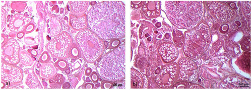
Histological sections of zebrafish ovaries, wild-type
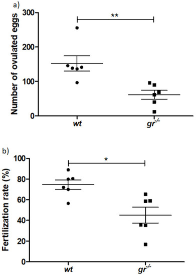
 (wt) (a), zebrafish glucocorticoid receptor (gr−/−) mutant line, nr3c1ia30/ia30 (gr−/−) (b). Eosin-Gill’s haematoxylin staining (10×). Scale bar 100 um.

Fish fertility (a) number of eggs laid by wt females crossed with wt males (black circle dots) and gr−/− females crossed with wt males (black square dots); (b) fertilization rate in wt and gr−/− females. Asterisks denote statistical significant differences (* p < 0.05; ** p < 0.01).

Infrared imaging analysis. (a) Microphotographs of class III oocytes in wt and gr−/− ovary sections. False color images showing the topographical distribution of: (b) lipids (color scale 0–6), (c) proteins (color scale 0–15), (d) glycosylated compounds (color scale 0–1), and (e) cortisol (color scale 0–8) (the dimensions of false color images were 328 × 328 mm). (f) Principal component analysis (PCA) scores’ plot of wt III (orange) and gr−/− (red) spectral data. (g) PC1 loadings of wt and gr−/− class III oocytes.

Hyperspectral imaging analysis. (a) Microphotographs of class IV oocytes isolated from wt and gr−/− ovaries; false color images showing the topographical distribution of (b) lipids (color scale 0–6), (c) proteins (color scale 0–15), (d) glycosylated compounds (color scale 0–1), and (e) cortisol (color scale 0–8) (the dimensions of false color images were 164 × 492 μm). PCA score plots of (f) wt IV (light green) and gr−/− IV (dark green) spectral data; PC1 loadings of (g) wt IV/gr−/− IV spectral populations.

Acknowledgments
This image is the copyrighted work of the attributed author or publisher, and ZFIN has permission only to display this image to its users. Additional permissions should be obtained from the applicable author or publisher of the image. Full text @ Int. J. Mol. Sci.