
- Title
-
Integrated Hypoxia Signaling and Oxidative Stress in Developmental Neurotoxicity of Benzo[a]Pyrene in Zebrafish Embryos
- Authors
- Lin, Y.C., Wu, C.Y., Hu, C.H., Pai, T.W., Chen, Y.R., Wang, W.D.
- Source
- Full text @ Antioxidants (Basel)

Developmental neurotoxicity of B[a]P revealed by the Tg(elavl3:EGFP) model. Tg(elavl3:EGFP) zebrafish embryos received the indicated treatments: 0 μM. (control, 01% DMSO; (A,C,F,I,L)), 10-μM B[a]P (B,D,G,J,M) and 20-μM B[a]P (C,E,H,K,N). (A–C) Lateral view of the live embryos at 24 hpf after treatment with 0.1% DMSO (control; A), 10-μM B[a]P (B) and 20-μM B[a]P (C); (D–O) Effects of B[a]P on neurogenesis using the Tg(elavl3:EGFP) model. The embryos treated with 0.1% DMSO (control; (D,G,J,M)), 10-μM B[a]P (E,H,K,N) and 20-μM B[a]P (F,I,L,O) were collected at 24 hpf. Representative fluorescent images are shown for embryonic trigeminal ganglions (tg) in the head region (D–F) and Rohon–Beard cell (rb) in the front trunk (G–I), mid-trunk (J–L) and in tail regions (M–O). (P–S) The numbers of tg cells in the head region and rb cells in front-trunk (F-trunk), mid-trunk (M-Trunk) and tail were counted from 10 embryos in each treatment group. Data are shown as mean ± standard deviation. * p < 0.05, ** p < 0.01, *** p < 0.001 compared with the control. |

B[a]P-treated embryos exhibit defective nervous system development. In situ hybridization staining was performed with a riboprobe against elavl3, a marker gene specifically expressed in telencephalon (te), hypothalamus (hy), midbrain (mb), midbrain hindbrain boundary (mhb) and plate (mhp) and hindbrain (hb) of the developing brain (A). (A–I) Zebrafish embryos exposed to 0 μM. (control, 01% DMSO), 10-μM B[a]P and 20-μM B[a]P were analyzed at 24 hpf. Embryos in (A–C) are shown from the lateral view; the red arrowheads indicate developmental defects in the midbrain (mb), and the hollow arrowheads indicate developmental defects in midbrain-hindbrain boundary (mhb); (D–F) Embryos are shown from the lateral view. The developing telencephalon (te; rectangles in (E,F)) was wider and the expression of elavl3 was weaker than in the control embryos; (G–I) Images are focused on the hindbrain (hb) region. The development of cranial ganglia (cg) was dose-dependently affected by B[a]P treatment; (J) Expression alteration of elavl3 in B[a]P was quantified by real-time quantitative RT–PCR (* p < 0.05, ** p < 0.01 compared with the control). |

Expression patterns of shh and isl1 are dose-dependently affected by B[a]P exposure. Wild-type (AB) zebrafish embryos were treated with 0 μM. (control, 01% DMSO), 10-μM B[a]P and 20-μM B[a]P. The embryos were fixed at 24 hpf. Expression of shh (A–G) and isl1 (H–M) was analyzed by in situ hybridization staining. Images show the lateral view in (A–C,H–J). Images in (E–G) show the dorsal view. Images in (K–M) show the frontal view; (B,C) Expression of shh was specifically expressed in the mid-diencephalic organizer (mdo), hypothalamus (hy) and basal plate (bp) of B[a]P-treated embryos; (E–G) The expression pattern of shh in the brain and floor plate (fp) in 20-μM B[a]P-treated embryos; (H–M) The development of encephalic regions was examined by in situ hybridization staining by probing for the islet1 gene (isl1) which is specifically expressed in telencephalon (te), ventral thalamus (vp), hypothalamus (hy), epiphysis (ep) and posterior commissure (pc) in embryos at 24 hpf. (N, O) Expression alteration of shhand isl1 in B[a]P was quantified by real-time quantitative RT–PCR (* p < 0.05, ** p < 0.01, ***p < 0.001 compared with the control). |

Lack of obvious neural cell defects in B[a]P-treated embryos at 12 hpf. In situ hybridization staining with a probe against elavl3 was performed to analyze the early development of neural cells (A–I); all images show the dorsal view. Images in (A–C) are focused on the anterior trunk; (D–F) are focused on mid-trunk region and (G–I) are focused on the tail region. tg, trigeminal ganglion; pm, primary motor neurons; rb, Rohon–Beard cells. |
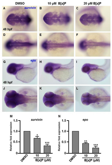
Expression levels of Survivin and Epo genes are reduced in B[a]P treated embryos. Zebrafish were exposed to 0 μM (control, 01% DMSO), 10 and 20 μM of B[a]P from 2 hpf until 24 hpf. Whole-mount in situ hybridization staining was performed with riboprobes against survivin (A–F) and epo (G–L) at 24 hpf. The staining levels of survivin (M) and epo (N) were quantified. * p < 0.05, ** p < 0.01, *** p < 0.001 compared to control. |
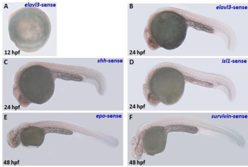
Sense riboprobe serves as negative controls for in situ hybridization staining assy. |