
|
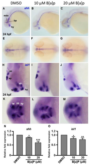
Fig. 3
Expression patterns of shh and isl1 are dose-dependently affected by B[a]P exposure. Wild-type (AB) zebrafish embryos were treated with 0 μM. (control, 01% DMSO), 10-μM B[a]P and 20-μM B[a]P. The embryos were fixed at 24 hpf. Expression of shh (A–G) and isl1 (H–M) was analyzed by in situ hybridization staining. Images show the lateral view in (A–C,H–J). Images in (E–G) show the dorsal view. Images in (K–M) show the frontal view; (B,C) Expression of shh was specifically expressed in the mid-diencephalic organizer (mdo), hypothalamus (hy) and basal plate (bp) of B[a]P-treated embryos; (E–G) The expression pattern of shh in the brain and floor plate (fp) in 20-μM B[a]P-treated embryos; (H–M) The development of encephalic regions was examined by in situ hybridization staining by probing for the islet1 gene (isl1) which is specifically expressed in telencephalon (te), ventral thalamus (vp), hypothalamus (hy), epiphysis (ep) and posterior commissure (pc) in embryos at 24 hpf. (N, O) Expression alteration of shhand isl1 in B[a]P was quantified by real-time quantitative RT–PCR (* p < 0.05, ** p < 0.01, ***p < 0.001 compared with the control).