FIGURE SUMMARY
Title

Zebrafish xenograft model of human lung cancer for studying the function of LINC00152 in cell proliferation and invasion

Authors
Shen, W., Pu, J., Sun, J., Tan, B., Wang, W., Wang, L., Cheng, J., Zuo, Y.
Source
Full text @ Cancer Cell Int.

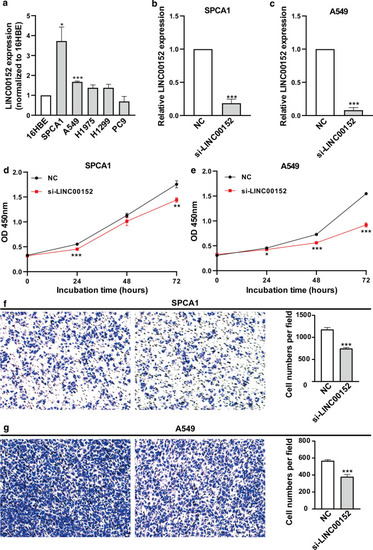
Knockdown of LINC00152 decreases the proliferation and invasion of lung cancer cells in vitro. a LINC00152 expression was analyzed by qRT-PCR in 5 NSCLC cell lines that were compared with the normal human bronchial epithelial cell line (16HBE). b, c LINC00152 mRNA levels in SPCA1 (b) and A549 (c) with LINC00152 siRNA (si-LINC00152) transfection compared with the negative control siRNA (NC) by qRT-PCR. d, e CCK-8 assays were performed to detect the viability of SPCA1 (d) and A549 (e) with LINC00152 siRNA transfection. f, g Transwell assays were performed to detect the invasion of SPCA1 (f) and A549 (g) with LINC00152 siRNA transfection. *: p < 0.05, **: p < 0.01, ***: p < 0.001

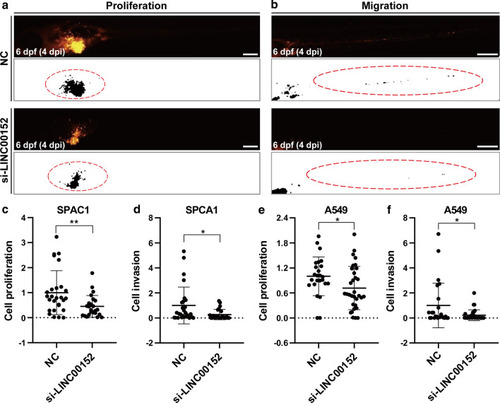
Knockdown of LINC00152 decreases the proliferation and invasion of lung cancer cells in zebrafish xenograft by stereomicroscopy. a, b Lung cancer cells transfected with si-LINC00152 siRNA or NC were injected into the PVS of 2-dpf WT zebrafish larvae. Images were taken using a stereomicroscope at 4 dpi. CM-DiI-positive areas in the yolk were quantified for proliferation (a), and CM-DiI-positive areas in trunk were quantified for invasion (b). The regions enclosed by the red dashed curve in the segmented images were selected for calculating tumor areas in the yolk or trunk. c, d Statistical analysis of proliferation (c) and invasion (d) when knocking down LINC00152 in SPCA1 cells. e, f Statistical analysis of proliferation (c) and invasion (d) when knocking down LINC00152 in A549 cells. *: p < 0.05, **: p < 0.01. Scale: 250 μm

Knockdown LINC00152 decreases the proliferation and invasion of lung cancer cells in zebrafish xenograft by confocal microscopy. a, b Lung cancer cells transfected with si-LINC00152 (a) or NC (b) were injected into the PVS of 2-dpf Tg(fli1a:EGFP) transgenic zebrafish larvae. Images were taken by confocal microscope at 4 dpi. Tumor areas in the yolk were quantified for proliferation, and the polygons enclosed by red dashed lines represent the tumor cell covering areas. The yellow arrow represents cell debris, which is excluded from the tumor cell covering areas. c, d Statistical analysis of proliferation (c) and migration (d) when knocking down LINC00152 in SPCA1 cells. e, f Statistical analysis of proliferation (e) and migration (f) when knocking down LINC00152 in A549 cells.*: p < 0.05, **: p < 0.01. Scale: 100 μm

Knocking down LINC00152 and blocking EGFR have a synergic effect on the inhibition of cell proliferation and invasion in vitro. a CCK-8 assays were performed to detect the viability of A549 with NC transfection, NC transfection and afatinib (EGFR inhibitor) administration, si-LINC00152 transfection, si-LINC00152 transfection and afatinib administration. b Transwell assays were used to investigate invasion with NC or si-LINC00152 transfection and afatinib administration or not. c Statistical analysis of invasion with NC or LINC00152 siRNA transfection and afatinib administration or not. ns, not statistically significant, **: p < 0.01, ***: p < 0.001

Knockdown of LINC00152 and block of EGFR have a synergic effect of inhibition in cell proliferation in zebrafish xenograft. a-d Lung cancer cells with NC transfection (a, b) or si-LINC00152 transfection were injected into the PVS of 2-dpf WT zebrafish larvae, and afatinib was bathed in zebrafish culture medium (b, d) or not (a, c). Images were taken by a confocal microscope at 3 dpt. e, f Statistical analysis of the proliferation and invasion with NC or LINC00152 siRNA transfection and afatinib administration or not. ns, not statistically significant, *: p < 0.05. Scale: 100 μm

Acknowledgments
This image is the copyrighted work of the attributed author or publisher, and ZFIN has permission only to display this image to its users. Additional permissions should be obtained from the applicable author or publisher of the image. Full text @ Cancer Cell Int.